In modern medicine, the intersection of digital imaging, additive manufacturing, and clinical expertise has given rise to a powerful tool that reshapes how surgeons approach complex operations. Three dimensional printing, also known as additive manufacturing, enables the creation of tangible, patient specific models from medical imaging data. These models provide a tactile representation of anatomy, pathology, and instrumentation pathways that can be held, examined from every angle, and manipulated before a patient is anesthetized. The impact extends beyond the mere novelty of a physical replica; it translates into clearer preoperative understanding, more precise surgical strategies, and the potential for improved patient outcomes. As teams gain experience, they integrate 3D printed designs into planning conferences, simulation sessions, and even the design of custom implants or surgical guides, progressively turning a digital plan into a concrete, actionable workflow. The role of 3D printing in surgical planning is thus best understood as a multi dimensional bridge linking radiology, anatomy, engineering, and the operating room, with patient safety and education as constants at the center of the process.
Origins and evolution of 3D printing in medicine
The roots of three dimensional printing date back to the late twentieth century, when early printers used sequential layering to transform digital geometries into physical objects. In medicine, pioneers recognized that accurately reproducing complex anatomical structures offered a unique advantage for understanding spatial relationships that are often difficult to appreciate on two dimensional scans. The initial applications were exploratory, focused on specific case reports and small series that demonstrated how a tangible model could reveal hidden recesses, deformities, or vessel relationships in areas such as the craniofacial skeleton and the skull base. As computing power grew and imaging modalities improved, the workflow matured; segmentation algorithms advanced to delineate tissues with greater precision, and printers evolved to handle a broader range of materials with improved surface finishes. Over time, academic centers and specialized manufacturers collaborated to build standardized pipelines that could produce reproducible models in a clinically meaningful timeframe. This evolution transformed 3D printing from a novel demonstration into a practical instrument that could be embedded within routine surgical planning and training.
With the broader dissemination of medical imaging datasets, more surgeons began to appreciate the value of patient specific models as a complement to traditional planning methods. The adoption was iterative: early experiences highlighted benefits in understanding complex anatomy, while later experiences underscored the potential to shorten operative times, reduce intraoperative uncertainty, and facilitate team communication. The evolution also reflected a shift from physical models alone toward integrated services that include custom surgical guides, templates, and, in some cases, end use implants designed to be sterilizable and compatible with sterile technique. Across specialties, the thread remains consistent: tangible representations of a patient’s anatomy help translate imaging into action, enabling a more informed and shared decision making process between clinicians and patients alike. This trajectory continues to be driven by advancements in scanning technologies, computer aided design, and novel printing materials that better mimic real tissue properties.
Technical foundations and workflow for surgical planning
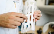
At the core of 3D printing in surgical planning is a carefully orchestrated workflow that converts clinical images into accurate physical objects. The journey typically begins with high quality imaging, most commonly from computed tomography or magnetic resonance imaging, sometimes complemented by specialized sequences or contrast materials. The data are then imported into segmentation software, where clinicians or trained technicians delineate structures of interest, such as bone, soft tissue, vessels, nerves, and tumors. The segmentation step requires meticulous attention to accuracy and may involve manual refinement after automated tools have performed initial contouring. The resulting three dimensional models are exported as standard file formats that preserve geometric fidelity, such as mesh representations that describe the surface of the structures. Once the digital model is ready, it is sent to a printer that uses a selected material system to build the object layer by layer, following the digital design. Before printing, considerations about sterility, biocompatibility, and mechanical properties guide the selection of materials and post processing steps, ensuring the final object meets the intended clinical use. Finally, the physical model is cleaned, cured, sterilized if necessary, and integrated into the surgical planning process alongside imaging, virtual simulations, and instrument selection. This end to end chain—from imaging to tangible model—forms a robust framework for translating data into actionable clinical insights.
Several printing technologies are commonly employed in surgical planning, each with its own strengths. Fused deposition modeling excels in producing durable, cost effective models that are suitable for education and rough planning. PolyJet or multi jet fusion techniques can deliver higher resolution surfaces and multi material capabilities, allowing a single model to showcase different tissue properties or to highlight critical structures with color coding. Stereolithography offers fine detail and smooth finishes that can be advantageous for delicate anatomical regions, though it may require careful post processing to achieve the desired mechanical characteristics. The choice of material is equally important: rigid plastics can faithfully reproduce bony architecture, while flexible polymers may mimic soft tissues. In some instances, combinations of materials and color coding help to convey spatial relationships more clearly, enabling surgeons to anticipate potential obstacles and to rehearse complex maneuvers before entering the operating room. The entire workflow demands cross disciplinary collaboration among radiologists, engineers, surgeons, and procurement specialists to ensure timely production, appropriate quality control, and accurate representation of patient anatomy.
Quality assurance is a critical component of this process. Each model should be validated for dimensional accuracy against reference imaging data, with verification steps that may include measurements, superimposition checks, or comparison against intraoperative findings. Documentation of the model’s parameters, printing workflow, and any post processing steps is essential not only for reproducibility but also for regulatory compliance and for informing future cases. As institutions accumulate experience, they develop internal standards that define acceptable tolerances, turnaround times, and material choices aligned with the planned surgical approach. Robust quality control helps to ensure that the model serves as a reliable surrogate for real anatomy and supports the decision making that precedes the operation. The integration of this pipeline into clinical practice rests on the perception that a well executed model reduces uncertainty, clarifies goals, and supports a safer, more efficient surgical plan.
Applications in different surgical specialties
Neurosurgery and skull base surgery have emerged as early beneficiaries of patient specific models. Complex cranial anatomy, with intricate relationships between tumor margins, vessels, and cranial nerves, benefits from tactile inspection and precise measurement of lesion extent. A physical model can guide tumor debulking strategies, help determine the feasibility of minimally invasive corridors, and support the planning of critical steps like bypass or skull base reconstruction. In craniofacial and maxillofacial procedures, models of congenital deformities or acquired trauma enable surgeons to rehearse osteotomies, plate bending, and implant placement with a level of foresight that reduces intraoperative guesswork. Orthopedic surgery leverages 3D printing to visualize deformities, plan corrective osteotomies, and even produce patient specific cutting guides that align precisely with the native anatomy. For complex joint reconstructions or spinal procedures, printouts and templates can aid in aligning hardware, optimizing nodal trajectories, and predicting the mechanical implications of implant choices. In cardiovascular surgery, models of congenital heart defects, aortic aneurysms, or intricate vascular networks support team based planning, device sizing, and the simulation of hemodynamic outcomes prior to intervention. Pediatric cases, where the anatomy may differ substantially from adult norms, particularly benefit from scaled models that help families understand proposed procedures and expected recoveries. Across these specialties, the common thread is that physical replicas translate imaging data into a shared, concrete reference that enhances planning accuracy and communication among the surgical team and with patients and families.
Dental and maxillofacial disciplines stand out for the seamless integration of 3D printed models with surgical guides and prosthetic components. In orthodontics, educational models complement digital simulations of tooth movement and occlusal schemes. In implant dentistry, guided surgery relies on precise drilling templates derived from patient scans, enabling accurate placement while minimizing invasiveness. The field of otolaryngology uses airway models to anticipate challenges during endoscopic or open procedures, and to train residents in delicate maneuvers without compromising patient safety. Vascular surgery benefits from arterial and venous replicas that enable the rehearsal of bypasses and aneurysm repairs, and occasionally, the designers create patient specific devices or clamps that conform to individual shapes. The expanding spectrum of applications reflects an underlying principle: when anatomy is made tangible, surgeons gain a more complete mental model, which translates into better plan quality and improved coordination of complex steps. This cross specialty versatility has driven the adoption of standardized workflows, shared repositories of case data, and opportunity for collaborative research across institutions.
Beyond direct operative planning, 3D printed models increasingly support simulation based training for residents and fellows. High fidelity trainers that reproduce tissue density and anatomical relationships allow learners to practice critical steps such as dissection planes, vessel preservation, and implant insertion in a risk free environment. Such simulations complement traditional curricula by providing objective practice opportunities and measurable performance benchmarks. In some centers, models are used in multidisciplinary conferences where radiologists, surgeons, anesthesiologists, and engineers discuss the case from a shared physical reference point, thereby aligning expectations and refining the surgical plan. This integrated approach helps to foster a culture of collaboration and continuous improvement, which is essential for maintaining high standards of patient care as techniques evolve and new devices enter the market.
Impact on preoperative planning and intraoperative guidance
The tangible impact of 3D printed models on preoperative planning extends across several dimensions. Clinicians report that models assist in visualizing relationships that are difficult to grasp on two dimensional images, enabling more precise measurement of lesion extent, bone thickness, and spatial orientation relative to critical structures. The ability to manipulate the model during planning sessions fosters a more collaborative dialogue among surgeons, radiologists, and support staff, leading to a shared mental representation of the case and a clearer consensus on the operative approach. In complicated reconstructions, models serve as a rehearsal platform where team members test different strategies, assess estimated blood loss, and anticipate potential complications. This preparatory work can reduce operative times, improve accuracy, and support safer transitions from planning to execution. Intraoperatively, the principles of planning are carried forward through the use of patient specific guides, templates, or implants that were designed using the same model system. For instance, a drilling guide designed from a printed bone replica can direct trajectories with a level of precision that translates to shorter anesthesia duration and minimized tissue disruption. The ability to translate a plan into a physical template reduces variance between what is intended and what is achieved during the operation, which is a meaningful contributor to surgical quality assurance.
Moreover, 3D printed models have proven valuable in enhancing patient communication and consent. When patients can see and touch a replica of their own anatomy, clinicians can explain the proposed procedure more clearly, discuss risks and benefits in relatable terms, and address anxieties more effectively. This aspect of patient centered care is increasingly recognized as a core component of informed consent, particularly in high stakes surgeries where understanding is essential for shared decision making. The educational payoff extends to students and trainees who gain a tangible sense of spatial relationships and technical considerations that are often difficult to convey through traditional images alone. In sum, the combination of improved planning precision, safer execution, and enhanced patient understanding represents a compelling argument for integrating 3D printing into comprehensive surgical workflows rather than treating it as a stand alone novelty.
Clinical outcomes associated with the use of 3D printed models are influenced by multiple factors, including case complexity, printing accuracy, and the overall experience of the surgical team. While evidence continues to grow, several patterns have emerged. In selected complex cases, models have correlated with shorter case durations, reduced need for intraoperative imaging, and more confident decision making in the face of anatomical surprises. In other settings, the benefits may be subtler but still meaningful, reflecting improvements in plan clarity, team coordination, and patient understanding. Importantly, the decision to employ 3D printing should be guided by a thoughtful assessment of expected benefits, timeline feasibility, and resource availability, rather than a universal, one size fits all approach. This nuanced use ensures that the technology remains aligned with clinical goals and patient safety while contributing to the ongoing advancement of surgical planning practices.
Finally, there is a growing recognition of the role of rapid prototyping and iteration in surgical design. In some cases, surgeons will generate multiple versions of a model to compare different treatment paths, such as variations in osteotomy angles or implant configurations. This iterative testing process can reveal subtle tradeoffs and help identify the most biologically compatible and technically feasible approach before any incision is made. The ability to iterate quickly and visualize outcomes fosters a culture of evidence based planning where decisions are grounded in tangible representations rather than abstract descriptions. As the field progresses, workflows increasingly embrace this iterative mindset, supported by improvements in software, materials science, and printing speed, which together create a powerful engine for refining surgical strategies and ultimately elevating patient care.
Educational value and patient communication
The educational dimension of 3D printing in surgery extends to both clinicians and patients. For trainees, physical models complement anatomical textbooks and virtual simulations by offering a hands on, tactile experience that reinforces anatomical knowledge, spatial reasoning, and procedural steps. This experiential learning can accelerate the development of proficiency in complex techniques, reduce the learning curve for challenging cases, and support the assessment of competency through hands on practice. For experienced surgeons, models provide a platform to experiment with novel approaches, rehearse cranial or facial reconstructions, and validate planning decisions against a tactile reference. In the realm of patient communication, 3D printed replicas help bridge gaps in understanding, enabling individuals to visualize their own anatomy, appreciate the scope of a proposed intervention, and participate more actively in decision making. This can lead to heightened satisfaction, improved trust, and a sense of collaboration with the care team, all of which contribute to the overall patient experience. Beyond individual consultations, models are increasingly used in public education campaigns, outreach programs, and multidisciplinary conferences where complex cases benefit from the clarity that a tangible object provides. The educational benefits thus propagate through the entire ecosystem of care, reinforcing the shared goal of safer, more effective surgery and enhanced patient engagement.
Training programs for residents and fellows are progressively integrating 3D printed models into curricula that emphasize competency based learning. Learners can practice critical steps such as exposure, dissection, and delicate maneuvers in a controlled environment that mirrors real anatomical challenges. By simulating different anatomical variations, instructors can expose trainees to a broader spectrum of clinical scenarios, building adaptability and problem solving skills. This exposure is particularly valuable in specialties that involve high stakes anatomy or rare conditions where hands on experience with actual patients is limited. Finally, the collaboration that stems from these activities fosters interdisciplinary communication, as radiologists provide accurate imaging representations and engineers contribute expertise on design and manufacturing aspects. The result is a richer educational experience that improves readiness for real world operations and supports the continual optimization of surgical practice.
Challenges, limitations, and safety considerations
Despite the promise of 3D printing in surgical planning, the field faces several challenges. One major consideration is the accuracy and fidelity of the printed model, which depend on image quality, segmentation precision, printer resolution, and material properties. Discrepancies between the model and the patient’s actual anatomy can lead to misleading conclusions if not carefully controlled. Time is another critical factor; generating, processing, and printing a model requires resources and coordination that must align with the clinical schedule, especially in urgent or emergency cases. Economic constraints also influence adoption, as institutions weigh the costs of equipment, materials, and personnel against anticipated benefits. In some settings, access to advanced printers and skilled technicians is limited, creating disparities in availability that may affect patient access to this technology. Sterilization and biocompatibility considerations are essential when models or guides are intended for intraoperative use. Some materials may tolerate sterilization processes poorly or may not mimic tissue properties closely enough for realistic simulation. Safety concerns extend to the potential for data privacy issues, particularly when patient imaging data are shared with outside vendors for model production. Rigorous data governance, informed consent, and compliance with regulatory standards are necessary to protect patient rights while enabling the benefits of physical modeling. Overall, the responsible integration of 3D printing requires thoughtful workflow design, ongoing quality assurance, and a transparent assessment of risk versus reward in each clinical scenario.
Another challenge centers on standardization. Across institutions, there can be variability in segmentation protocols, material choices, and printing parameters, which can complicate multi center studies and comparisons of outcomes. Efforts to establish consensus guidelines and shared best practices are essential to ensure reproducibility and reliability of models as evidence of clinical value. As technology advances, newer materials, higher resolution printers, and advanced software algorithms continuously reshape what is possible, necessitating periodic reevaluation of workflows and training. Clinicians must remain vigilant about the limits of the technology, avoiding over reliance on models for decisions that require dynamic physiological assessment or intraoperative flexibility. A balanced approach that integrates 3D printing with other planning modalities ensures that patient safety remains paramount while still leveraging the benefits of tangible representation and simulation. The safety landscape thus requires ongoing collaboration among surgeons, engineers, regulators, and manufacturers to align capabilities with patient needs and ethical considerations.
Regulatory, ethical, and data management aspects
Regulatory considerations for 3D printed surgical models and related devices vary by jurisdiction but share common themes centered on safety, efficacy, and quality control. Clinicians and manufacturers must adhere to medical device regulations when the model is used for patient care in a way that could influence surgical decisions or when a component like a guide or custom implant is created for clinical use. Documentation, traceability, validation, and post market surveillance are integral parts of responsible practice. Ethical considerations include ensuring informed consent for the use of personalized data and clear communication about the potential benefits and limitations of 3D printed planning tools. Transparency about resource allocation, potential biases, and the learning curve associated with adopting the technology helps maintain trust with patients and the medical community. Data management is another critical axis, given that imaging data are sensitive personal information. Secure data handling, de identification when appropriate, and careful control of access to files and printers are necessary to protect privacy while enabling collaboration with multidisciplinary teams and external partners. The convergence of technology, medicine, and law creates a landscape where careful governance supports innovation while safeguarding patient welfare and professional integrity.
Beyond regulatory and ethical concerns, the sustainability of 3D printing programs depends on thoughtful assessment of long term value. Institutions may evaluate metrics such as time saved in planning, reductions in intraoperative complications, or improvements in patient comprehension and satisfaction. These measures help determine whether investments in printers, materials, and personnel yield meaningful clinical return. Collaboration with industry partners can accelerate access to cutting edge materials and printing modalities, but it is essential to maintain rigorous oversight to prevent conflicts of interest and protect clinical objectivity. Building a culture of continuous improvement, ongoing training, and strict quality assurance ensures that 3D printing remains a safe, effective adjunct to surgical planning rather than a stand alone technology. In this sense, the role of governance is as important as the engineering itself, guiding responsible growth and ensuring that patient well being remains the central priority while exploration and innovation continue unabated.
Future directions and ongoing research
The horizon for 3D printing in surgical planning is broad and dynamic, driven by advances in imaging, materials science, computational modeling, and data integration. Innovations in multi material printing allow the creation of models that simulate distinct tissue properties, such as stiffness and texture, to provide more realistic tactile feedback during planning and simulation. Developments in rapid prototyping and on demand manufacturing aim to shorten turnaround times, enabling near real time planning for select procedures and expanding access in urgent settings. The integration of augmented reality and virtual reality with physical models may offer hybrid planning environments where surgeons can switch seamlessly between tangible replicas and immersive digital overlays to rehearse complex maneuvers. Advances in automation and artificial intelligence promise to streamline segmentation, improve accuracy, and reduce the time required to produce high quality models, making these tools more accessible to a wider range of institutions. In clinical research, standardized reporting of outcomes related to 3D printed planning will help practitioners draw robust conclusions about the technology’s impact on safety, efficacy, and efficiency. The continued collaboration among radiologists, engineers, material scientists, and surgeons will drive iterative improvements that align the capabilities of 3D printing with the nuanced demands of diverse surgical specialties. As the field evolves, the role of 3D printing in surgical planning is likely to expand from a supportive aid to a central component of personalized, precision driven care that harmonizes imaging data, manual skills, and patient outcomes in a cohesive, predictable workflow.