Introduction to 3D printing in modern healthcare
In the unfolding story of modern medicine, the advent of three dimensional printing has emerged as a transformative force, reshaping how clinicians approach diagnosis, planning, intervention, and postprocedural recovery. This technology, which began as a method for rapid prototyping in industrial settings, has evolved into a versatile platform capable of translating complex digital models into tangible objects with remarkable precision. Its impact on medicine arises from the unique ability to tailor tools, implants, and educational materials to the individual patient, bridging the gap between theoretical design and real world application. The result is a shift toward personalization, a reduction in procedural uncertainty, and a potential to shorten the path from concept to clinical impact. The early uses were often experimental, driven by engineers collaborating with surgeons, radiologists, and researchers who recognized that a physical replica of an organ or a defect could illuminate anatomy in ways that static images could not. Over time, as imaging resolution improved and software became more accessible, 3D printing matured into a practical component of routine care, trialed in planning rooms, operating suites, dental clinics, and research laboratories around the world. The broad spectrum of applications has continued to expand, guided by a growing body of evidence and a willingness to explore new materials, new workflows, and new regulatory pathways that ensure safety and efficacy. In contemporary practice, the role of 3D printing is defined not by a single breakthrough but by a continuum of innovations that collectively empower clinicians to visualize, simulate, and customize medical solutions with an unprecedented level of detail and personal relevance.
Across specialties, the technology is often described as a bridge between digital data and physical reality. Medical imaging provides rich, patient specific information about anatomy and pathology, and 3D printing converts this information into objects that can be held, examined, and manipulated. The tactile dimension added by printing complements the visual cues from scans, enabling more precise planning and communication among care teams, as well as between clinicians and patients. This capacity to produce patient specific models contributes to a deeper understanding of complex anatomies, facilitates shared decision making, and allows for risk assessment in ways that static models cannot replicate. The clinical impact extends beyond the operating room; in education and training, students and residents gain hands on experience with realistic replicas that reinforce learning, foster procedural confidence, and reduce error potential. The evolving ecosystem of devices, materials, and workflows paints a picture of a healthcare landscape where customization, speed, and accuracy are not competing priorities but converging objectives that advance patient outcomes across settings and populations.
Technologies and materials enabling medical 3D printing
The landscape of 3D printing technologies used in medicine encompasses a range of processes, each with distinct strengths and limitations that shape their suitability for specific clinical tasks. Fused deposition modeling, commonly known as FDM, builds parts layer by layer from thermoplastic polymers and has the advantages of simplicity, speed, and relatively low cost. Stereolithography, or SLA, utilizes light to cure photosensitive resins, delivering high resolution and smooth surface finishes that are particularly valuable for detailed anatomical models and surgical guides. Selective laser sintering, often referred to as SLS, employs a laser to fuse powdered materials, enabling durable parts with good mechanical properties and the capacity to produce complex geometries without extensive support structures. PolyJet and multi jet modeling extend these capabilities by depositing multiple materials and colors in a single print, offering a spectrum of stiffness, translucency, and biocompatible options that can faithfully simulate tissues of differing properties. Metals such as titanium are used in implants and implants related tooling when strength and biocompatibility are paramount, while ceramics and bioinert polymers serve in areas where chemical stability and sterilization compatibility are critical. The selection of material is guided not only by mechanical requirements but also by sterilization pathways, regulatory constraints, and compatibility with imaging modalities used in the clinical workflow. In parallel with structural materials, advances in bioprinting introduce hydrogel based bioinks and composite formulations designed to support cell viability and tissue maturation in research settings, with the potential to translate into regenerative therapies as science and manufacturing scale align. The convergence of these technologies with robust software ecosystems that enable precise segmentation, design, and verification underpins the reliability of printed medical items, and the trajectory of this field continues to be shaped by ongoing innovations in materials science, process engineering, and quality assurance frameworks that must keep pace with clinical demands. In practice, this means that teams must select printing modalities that balance accuracy, speed, cost, and biocompatibility while maintaining strict control of variables that impact sterility, reproducibility, and patient safety. The careful integration of sterilization protocols, post processing steps, and validation testing is essential to translate a digital concept into a clinically usable object that can withstand the rigors of use in the human body or in the operating environment.
In addition to the conventional plastics and metals, the medical 3D printing field increasingly embraces composite materials and functional additives that can imbue printed parts with specific properties such as radiopacity, color coding for anatomical labeling, and even drug elution capabilities in specialized devices. The challenge of integrating these complex materials lies not only in achieving the desired mechanical performance but also in ensuring that the printing process itself does not degrade material integrity or introduce contaminants. Consequently, interdisciplinary collaboration among engineers, material scientists, clinicians, and regulatory specialists becomes essential to design, test, and implement print processes that consistently deliver safe products. The regulatory landscape, while evolving, emphasizes traceability, documentation, and rigorous quality control, reminding practitioners that the promise of customization must be balanced with the strict standards that govern patient care. As the technology matures, the field is also learning how to manage the lifecycle of printed devices—from digital file stewardship and version control to field repair and end of life disposal—so that innovation remains responsible and sustainable while expanding access to high quality solutions. The combined effect of these technologies and materials is a growing toolbox that clinicians can draw upon to tailor interventions to each patient, to visualize complex procedures, and to train with equipment that mirrors real world scenarios in a safe and controlled manner.
From imaging data to tangible objects: the workflow
At the core of medical 3D printing is a workflow that begins with high quality imaging data, typically acquired through computed tomography or magnetic resonance imaging. The first phase involves careful data handling to preserve the fidelity of anatomical structures while enabling subsequent manipulation in a digital environment. Skilled technicians and clinicians work together to segment the imaging data, isolating bones, soft tissues, vessels, or pathological features that will become the basis for a printable model. The segmentation process translates grayscale image data into a three dimensional representation that can be refined using computer aided design software. Once a digital model is constructed, engineers and clinicians collaborate to adjust surface topology, ensure appropriate wall thickness, and incorporate any features that will be needed for surgical planning, simulation, or manufacturing. The model then undergoes verification steps to confirm that dimensions correspond to the patient’s anatomy within clinically acceptable tolerances. The final stage involves selecting a printing modality and material that satisfy the intended use; the file is prepared with attention to print orientation, support structures, and post processing requirements to optimize strength and surface finish. After printing, parts undergo cleaning, curing, or sterilization as applicable, followed by inspection for dimensional accuracy and integrity before they are used in a clinical setting. This workflow is not static; it is continuously refined through feedback from surgeons, radiologists, and therapists who rely on printed objects to augment their decision making or to simulate realities that cannot be captured with imaging alone. The digital to physical translation thus becomes a collaborative process that blends technical precision with clinical insight, converging on solutions that enhance patient safety, reduce uncertainty, and improve the overall quality of care.
Clinical applications: planning, guidance, and training
In the hands of skilled practitioners, patient specific models act as a bridge between imaging and intervention, enabling more informed planning of complex procedures. For example, in craniofacial reconstruction or intricate orthopedic corrections, a tangible replica of a patient’s anatomy allows surgeons to rehearse the steps of a challenging operation, identify potential difficulties, and tailor the approach to the contours of the individual bone and soft tissue envelope. This tangible rehearsal can shorten operative times, refine instrument selection, and improve intraoperative orientation, all factors that contribute to better outcomes and safer anesthesia management. For trainees, access to high fidelity replicas of human anatomy provides an invaluable level of experiential learning that complements textbooks and cadaveric work, thereby accelerating skill acquisition and confidence. Beyond the operating room, printed models facilitate patient education by offering a concrete visualization of the condition and the proposed treatment, often easing complex conversations and supporting informed consent. In oncology, printed representations of tumors and surrounding critical structures can illuminate the spatial relationships that influence resection margins, nerve preservation, and organ function, assisting multidisciplinary teams in forming consensus on the optimal surgical plan. Moreover, models are increasingly used to plan radiation therapy, where the precise geometry of the target and critical structures must be integrated with dose calculations to minimize collateral damage. In all these scenarios, the interplay of precise geometry, material realism, and tractable workflow creates a powerful toolkit that enhances precision medicine and patient centered care while reinforcing the culture of meticulous planning that characterizes high quality clinical practice.
Training environments also benefit from anatomically accurate synthetic replicas that can withstand repeated use, allowing clinicians to simulate not only the mechanics of a procedure but also the tactile feedback that guides instrument handling. The tactile dimension helps trainees build muscle memory in a controlled setting and fosters a safety oriented mindset when transitioning to real patients. This educational emphasis dovetails with research oriented activities, where printed prototypes are used to test new devices, explore design iterations, and validate computational models before moving into animal or human studies. The cumulative effect of these uses is a more efficient workflow in which risk is identified early, decisions are better informed, and the patient experience is enhanced by clearer communication and more consistent care pathways. As printing technologies continue to evolve together with imaging modalities and surgical techniques, the scope and fidelity of printed planning tools are likely to grow, enabling increasingly sophisticated simulations and more nuanced operational strategies that can adapt to the unique anatomy of each patient.
Implants, prosthetics, and personalized devices
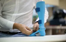
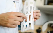
Printed implants and prosthetic components embody the most direct translation of additive manufacturing into lasting clinical infrastructure. The ability to tailor implants to patient specific anatomy has led to more natural fit, improved integration with biological tissues, and, in some cases, faster recovery trajectories. Cranial implants produced with biocompatible polymers or titanium alloys can be designed to complement the exact contours of a skull defect, offering improved aesthetic and functional outcomes while reducing the need for intraoperative contouring that can extend surgery time. Dental prosthetics and orthodontic appliances pursue similar goals, delivering precise occlusal relationships and improved patient comfort. In the realm of orthopedics, patient specific guides for bone cuts, drilling templates, and custom plates reduce intraoperative guesswork and can standardize complex procedures across surgeons with varying levels of experience. In prosthetics for limbs or facial features, patients may benefit from sockets, sockets liners, or externally attachable components that conform to residual limb geometry and gait requirements, thereby improving energy efficiency and comfort over long term use. While these advances are transformative, they also raise important considerations about sterilization, biocompatibility, supply chain reliability, and the need for durable validation under real world conditions. The ongoing collaboration among engineers, surgeons, and regulatory experts is essential to ensure that personalization does not compromise safety or long term performance, and that clinicians can anticipate how printed devices behave throughout the patient’s life cycle.
Pharmaceutical and drug delivery innovations
3D printing has begun to reshape pharmaceutical development and patient centered therapy by enabling dose customization, combination therapies, and targeted delivery mechanisms that align with individual treatment needs. The ability to fabricate oral dose forms with complex release profiles allows clinicians to tailor the pharmacokinetics to a patient’s physiology and comorbidities, potentially improving therapeutic outcomes while reducing adverse effects. A landmark achievement in this area was the approval and subsequent use of 3D printed tablets that demonstrate flexible release characteristics, illustrating how additive manufacturing can accommodate personalized regimens beyond the capabilities of conventional manufacturing. In addition to oral dosage forms, research explores implantable drug delivery devices and transdermal systems, where micro scale channels and reservoirs embedded in biocompatible matrices can regulate drug release with high precision. The regulatory environment in this domain emphasizes quality control, batch traceability, and robust stability testing, highlighting the need for rigorous manufacturing standards and post market surveillance. Clinicians, pharmacists, and biomedical engineers collaborate to translate these innovations from laboratories into clinical protocols, ensuring patient safety while expanding therapeutic options. As this field matures, the convergence of patient specific needs with materials science and pharmacology holds the promise of therapies that are not only effective but also tailored to individuals at the level of precise dosing, timing, and delivery route. Yet this promise is balanced by the responsibility to maintain stringent controls over manufacturing variability, to thoroughly characterize interactions with human biology, and to remain steadfast in ethical considerations about access and equity in treatment options.
Education, research, and simulation in medicine
The educational dimension of 3D printing in medicine extends beyond patient care into the realms of science and innovation, fueling a virtuous cycle of learning, experimentation, and dissemination. In medical education, realistically contoured models of organs, vasculature, and pathologies enable students and residents to study anatomy in three dimensions with a level of immersion unattainable from textbooks alone. Simulation environments equipped with printed anatomical replicas allow teams to practice complex procedures, refine communication protocols, and rehearse contingency plans in a low risk setting. This experiential learning translates into greater procedural confidence and improved team performance when real cases arise. Researchers leverage printed components as rapid prototyping tools for new devices, enabling iterative testing and design optimization before committing to expensive or time consuming manufacturing processes. In translational science, printed scaffolds and organ like constructs provide tangible platforms to study tissue response, mechanical behavior, and integration with host tissues, bridging the gap between computational predictions and biological realities. The dissemination of open access models and collaborative design repositories accelerates knowledge sharing, empowering institutions with limited resources to participate in cutting edge developments. As education and research continue to intertwine with clinical practice, 3D printing becomes a universal language through which theory is tested, validated, and refined in service of better patient care and scientific advancement. The ethical dimension of education remains central, as instructors ensure accuracy, consent, and appropriate use of models while students cultivate competencies that emphasize patient safety and professional responsibility.
Regulatory, safety, and ethical considerations
The integration of 3D printing into medicine is governed by a framework that seeks to balance innovation with patient protection, quality assurance, and accountability. Regulatory bodies across jurisdictions emphasize a rigorous demonstration of safety, efficacy, and reproducibility for printed medical devices and patient specific tools that interact with the human body or support surgical decisions. This includes documentation of material biocompatibility, sterilization validation, and software validation for digital workflows, all of which contribute to reliable, traceable, and auditable manufacturing processes. Quality management systems, process controls, and post production release criteria are essential to ensure that each printed item meets defined standards before clinical use, and this is complemented by ongoing surveillance to detect drift and to address emerging risks. Ethical considerations focus on patient autonomy, informed consent, and equity in access to printed solutions, particularly as cost or resource constraints could influence availability in underserved communities. Privacy concerns arise when digital medical data are used to create physical models, underscoring the necessity of secure data handling and proper deidentification when appropriate. The global landscape features a spectrum of regulatory maturity, with some regions adopting considerably stringent guidelines and others balancing innovation incentives with safeguard measures. Clinicians and institutions must navigate this landscape with transparent communication, rigorous validation, and a shared commitment to patient welfare. In practice, successful implementation of 3D printing in medicine requires cohesive collaboration among surgeons, engineers, radiologists, administrators, and regulatory affairs professionals who together develop evidence that justifies clinical use, demonstrates benefits, and clarifies limitations so that patients receive care that is both advanced and responsibly delivered.
Economic impact, accessibility, and future prospects
From a health systems perspective, the economics of 3D printing in medicine involve analyzing upfront investment, operating costs, and the downstream effects on clinical outcomes, workflow efficiency, and patient experience. While initial equipment purchases and training represent significant expenditures, the long term potential includes reductions in operative time, decreased reliance on outsourcing for models and guides, and improved resource utilization through more precise planning. In settings where access to specialized implants or housing materials is limited, in house printing and digital fabrication can enable timely interventions that would otherwise be delayed by procurement bottlenecks, translating into shorter hospital stays and faster rehabilitation. Cost variability is influenced by material choice, device complexity, scale of production, and the required regulatory oversight, which in turn shapes reimbursement strategies and incentives for adoption. Accessibility is also impacted by the availability of skilled personnel, the strength of digital infrastructure, and the capacity to implement robust quality assurance and sterilization processes. Looking forward, advances in open design ecosystems, cloud based collaboration, and standardized validation protocols hold promise for broader dissemination of 3D printing capabilities without compromising safety or consistency. The convergence of automation, artificial intelligence driven design, and multi material printing could unlock a future where patient specific solutions are created quickly, delivered reliably, and integrated into routine care with predictable costs. At the same time, ethical considerations about equitable access, intellectual property, and environmental impact must be addressed as the technology becomes more widespread, ensuring that the benefits of 3D printing in medicine reach diverse populations and do not exacerbate existing disparities in healthcare delivery. The trajectory points toward a healthcare environment where teams routinely incorporate printed models and components into the therapeutic plan, where patient engagement is enhanced by tangible demonstrations, and where innovation translates into measurable improvements in safety, effectiveness, and quality of life for patients facing complex medical challenges.