Overview of the Technology and Its Emergence in Cardiology
Three dimensional printing, also known as additive manufacturing, has evolved from a prototyping curiosity into a mature clinical tool across many medical specialties, with cardiology standing out as an area where tangible models can translate complex imaging data into tactile understanding. In its essence, this technology builds objects layer by layer from digital designs, and in medicine it has moved beyond simple plastics into biocompatible materials that can mimic the mechanical properties of human tissues. The heart, with its intricate geometry, variable wall thickness, and dynamic motion, presents unique challenges that digital representations alone cannot satisfy. With the advent of high resolution imaging, including computed tomography and magnetic resonance imaging, clinicians gained access to highly detailed anatomical datasets that can be converted into physical replicas. The real value emerges when a patient-specific model, created from the exact anatomy of an individual, can be held, rotated, cut, and examined from angles that would be difficult to visualize with two dimensional projections alone. This tangible interface allows surgeons and interventionalists to rehearse complex procedures, weigh alternative strategies, and communicate with patients in a more concrete way, thereby bridging the gap between imaging interpretation and real world execution. The trajectory of adoption has been shaped by advances in software for segmentation and meshing, improvements in printer hardware, and the refinement of materials that simulate not only the shape but also the texture and compliance of cardiac tissues. The result is a spectrum of models that can range from rigid anatomical replicas to flexible, elastic structures that respond to pressure, thereby enabling functional simulations that echo physiological realities. In addition to the clinical dimension, there is an educational and research component that is equally compelling, as medical students, fellows, and engineers can study heart anatomy across a continuum of pathologies with a consistent and reproducible tactile reference. The early enthusiasm gave way to a more measured but steadily expanding practice, supported by growing evidence that patient-specific models can influence decision making, reduce procedure time, and improve communication within multidisciplinary teams. As the field matures further, standardization of workflows, validation against surgical outcomes, and thoughtful integration into clinical pathways will determine how deeply 3D printing reshapes the standard of care in cardiology while maintaining patient safety, cost effectiveness, and ethical stewardship of resources.
The technology rests on three pillars that interlock to produce clinically meaningful outputs. First is accurate data acquisition, where high fidelity imaging captures the anatomy of interest with sufficient resolution to delineate delicate structures such as valve leaflets, annular dimensions, and the fine vasculature around the heart. Second is robust data processing, including segmentation to isolate relevant structures, surface generation that cleanly represents borders, and often the creation of a printable mesh that honors both external contours and internal cavities. Third is material selection and printing technology, which determine how closely a model can mimic tissue properties, how durable it is for repeated handling, and how cost effective the production is for routine clinical use. The interplay of these steps dictates the degree to which a model can serve as an accurate stand in for the living organ and as a reliable platform for predicting how interventions will unfold in real patient scenarios. In clinical practice, the ideal model is both anatomically precise and mechanically representative, a combination that enables rehearsals that are not merely aesthetic but kinetically informative. As materials science advances, clinicians gain access to polymers and composites that approximate myocardium, blood vessel walls, and calcified deposits with varying stiffness and response to deformation. The field continues to explore the balance between realism and practicality, recognizing that sometimes a highly accurate geometric replica is sufficient for planning, while in other cases a dynamic, flexible model that can be inflated or pressurized to simulate cardiac cycles becomes indispensable for understanding function under load.
From Imaging to Model: The Cardiac 3D Printing Workflow
The journey from medical image to a physical cardiac model begins with data acquisition, where the quality and consistency of the source imaging set the stage for successful downstream processing. In cardiology, CT angiography offers exceptional spatial detail of coronary anatomy and cardiac chambers, while MRI provides soft tissue contrast and functional information that can be crucial for understanding dynamic processes. Echocardiography, particularly transesophageal and three dimensional modes, contributes real time insights into valve motion and chamber relationships. The choice of imaging modality depends on the clinical question, and in many cases a combination of datasets is fused to create a comprehensive representation of anatomy. Once imaging data are secured, the next phase is segmentation, a computational step in which the region of interest is delineated from surrounding structures. This task can be performed with specialized software that allows semi-automatic detection of interfaces between blood, myocardium, valvular tissue, and vasculature. Segmentation accuracy is affected by image resolution, artifacts, and the inherent variability of biological tissue, making meticulous review by an experienced operator essential to prevent propagation of errors into the final model. After segmentation, a digital model is generated, often in the form of a stereolithography or mesh file, which then undergoes a process called meshing. This step ensures the surface geometry is clean, watertight, and suitable for printing, and may include smoothing, hollowing of chambers, or creating channels to represent vessels. For models intended to behave like living tissue, walls may be assigned specific thicknesses or flattened sections to facilitate printing with a chosen material. The subsequent step involves selecting the printing technology and material system that best align with the clinical aim. Polymeric resins, photopolymers, thermoplastics, and elastomeric materials each offer distinct mechanical profiles, ranging from rigid exact replicas to flexible, compliant structures that better simulate the pliability of cardiac tissue. For some applications, multi material printing enables simultaneous representation of different tissue types within a single model, such as a rigid calcified valve annulus adjoining a softer ventricular wall. After printing, models may require post processing, including curing, cleaning, and sometimes surface finishing to improve tactile feel or to reduce porosity that could trap air pockets. In addition to the physical model, some workflows embed sensors or channels to enable functional testing or to observe the interaction of devices with the anatomy during a bench simulation. Finally, the model is validated by comparing measurements and geometry against the original imaging data and, crucially, by consulting the clinical context to determine whether the model will meaningfully inform a particular decision, be it planning a surgical approach, selecting a device, or communicating risk to a patient. The cumulative workflow, while technically demanding, is increasingly guided by standardized protocols and institutional experience, which helps ensure reliability and reproducibility across cases.
In practice, clinicians emphasize that a well-designed cardiac model should offer a closed representation of necessary features without becoming an overfitted homage to imaging data. A balance must be struck between fidelity and practicality, recognizing that some simplifications may be permissible when they retain the essential anatomical relationships that govern procedural strategy. The printing phase itself has evolved with advances in desktop and industrial printers alike, expanding the accessibility of this technology across hospital centers and regional networks. While high-end workstations and professional grade printers provide exceptional resolution and material options, more economical printers still yield meaningful results for teaching, preoperative planning, and device testing. The choice of materials is particularly critical in simulations of blood flow, where compliant elastomeric materials may be paired with transparent shells to observe contrast flow or to simulate catheter advancement. In other scenarios, rigid materials with high dimensional stability can faithfully reproduce calcified structures, allowing practitioners to anticipate potential interactions between devices and hard tissue. As practitioners gain experience with these workflows, there is growing emphasis on documenting the end-to-end process, including imaging parameters, segmentation decisions, model dimensions, printing settings, and any post processing that could impact measurements. Transparent reporting not only supports quality control but also promotes learning across teams by making the rationale behind decisions explicit and reproducible for future cases.
Clinical validation of 3D printed models remains an evolving domain, with studies exploring how tactile models influence surgical planning time, choice of intervention, and surgeon confidence. Observational reports and small trials have highlighted benefits such as enhanced communication with patients and families, improved understanding of complex anatomies, and the potential for reducing intraoperative surprises. Yet, researchers also acknowledge limitations, including the time and cost associated with producing a model, the need for specialized personnel to manage segmentation and printing, and the risk of overstating the predictive value of a static replica in a dynamic physiological environment. As multi disciplinary teams refine their approaches, more rigorous comparative studies, standardized metrics for model accuracy, and economic analyses will help determine the most efficient ways to integrate 3D printing into daily cardiology practice. In parallel, emerging technologies, such as augmented reality overlays and virtual reality simulations, may complement physical models by providing immersive visualization that can be coupled with haptic feedback, creating a more holistic training and planning environment. The future workflow could envision an integrated platform where imaging, simulation, printing, and post-procedure evaluation feed into a continuous loop that advances both clinical skills and patient outcomes.
Clinical Applications in Cardiology
One of the most visible domains where 3D printing has made an impact is congenital heart disease, where the anatomy can be exceptionally complex and patient-specific. For children and adults with congenital defects, reconstructive strategies often hinge on precise understanding of spatial relationships among chambers, great vessels, valves, and outflow tracts. A patient-specific model can illuminate subtle nuances such as the orientation of a ventricular septal defect in relation to the conduction system, the course of anomalous coronary arteries, or the spatial constraints posed by a malpositioned ventricle. By physically manipulating the model, surgeons and interventionalists can anticipate potential challenges, test alternative routes for catheter access, and practice suturing or patching techniques before entering the operating room. This hands-on rehearsal translates into greater confidence, reduced ischemic time, and sometimes shorter overall procedure durations, all of which can contribute to improved patient safety and more efficient use of operating room resources. Beyond planning, these models serve as valuable tools for family discussions, helping caregivers grasp the intricacies of the condition and the intended therapeutic plan with a tangible reference that complements traditional imaging narratives. In addition to congenital heart disease, 3D printed models are increasingly used for complex valvular disease, including cases with multiple regurgitant streams or unusual annular geometries where valve repair carries substantial technical demands. Surgeons can evaluate the fit and mobility of a prosthetic or repair technique against the exact topography of the patient’s anatomy, and interventional teams can rehearse transcatheter procedures with a high degree of realism before committing devices to a live patient. The ability to simulate device deployment in a patient-specific context helps in risk stratification and may influence device selection, sizing, and approach, particularly in anatomies that deviate from textbook norms. In the realm of coronary artery disease, 3D printed models help illuminate the three dimensional pathways of branches, the relationship of plaques to vessel walls, and the spatial challenges encountered during complex interventions such as left main artery bifurcation stenting or chronic total occlusion procedures. Clinicians can use these models to discuss potential strategies for revascularization, visualize the proximity of lesion sites to critical structures, and plan the sequence of device deliveries in a controlled, reproducible setting. For electrophysiology, models that incorporate pacing leads, scar tissue, and conduction pathways enable better planning of ablation strategies and device positioning, enhancing both the safety and efficacy of interventions. Across these domains, the common thread is that tangible representations translate plan into practice in ways that radiographic slices and digital renderings sometimes cannot fully convey, thereby enriching the diagnostic and therapeutic landscape of modern cardiology.
In addition to procedures, 3D printed models have found a role in device development and testing. Engineers and clinicians collaborate to validate new valve designs, left atrial appendage closure devices, or novel catheters in anatomically faithful replicas before moving into animal studies or early human trials. This translational bridge accelerates innovation, as feedback from realistic simulations can inform design refinements, reduce iterative cycles, and support regulatory submissions with practical demonstrations of how a device interacts with complex anatomy. The iterative nature of this work, coupled with robust documentation, can shorten timelines for bringing safer, more effective devices to patients. It is important to remember that while physical models are powerful aids, they complement rather than replace rigorous clinical evaluation, and the integration of 3D printing into research pipelines should be guided by sound scientific principles, appropriate controls, and clear endpoints that reflect meaningful clinical outcomes. The broad spectrum of applications—from planning and education to device development—reflects a growing recognition that tactile, patient-specific understanding of heart anatomy can augment the standard repertoire of imaging modalities, enhancing both clinician performance and patient engagement in the care journey.
In medical education, 3D printed cardiac models serve as compelling tools for training learners at different levels, from medical students to seasoned fellows. The ability to handle specimens that replicate normal and pathological anatomy fosters an experiential understanding that complements theoretical instruction. Students can identify landmarks, measure spatial relationships, and practice basic technical skills such as suturing or catheter navigation in a controlled setting that resembles clinical reality. For trainees, repeated interaction with physically accurate replicas reinforces spatial memory and procedural sequencing, which can reduce cognitive load during actual procedures. In addition to clinical education, these models support multidisciplinary team training, where surgeons, interventional cardiologists, anesthesiologists, perfusionists, and nursing staff rehearse complex cases together. Such rehearsals promote communication, clarify roles, and help synchronize team performance under the high stakes conditions typical of cardiac procedures. The educational advantages extend to patient education as well, because patients and families often respond positively to seeing and handling a replica of the heart that corresponds with the information explained during consultations. This experiential learning can demystify disease processes and contribute to more informed consent, better expectation management, and a sense of shared decision making. Across settings, the educational impact of 3D printing aligns with broader goals of medical training, including standardized practice, competency-based progression, and the cultivation of confidence through deliberate exposure to realistic anatomical scenarios.
Ethical considerations accompany the clinical and educational benefits of 3D printing. Access and equity must be addressed to prevent disparities in who benefits from this technology, particularly given the upfront costs and the need for specialized expertise. Institutions must balance the potential improvements in patient care with prudent stewardship of finite resources, ensuring that models are used where they provide tangible value and not simply as a novelty. Informed consent processes should explicitly address the use of patient imaging to generate physical models, including privacy protections and the intended purpose of the models within the care plan. When models influence procedural decisions, ongoing evaluation should be performed to verify that benefits materialize in patient outcomes and are not solely demonstrable in theoretical discussions or planning sessions. Sustainable practice also calls for ongoing assessment of materials and equipment to minimize waste and to identify recyclable or reusable components where possible. As the field expands, regulatory frameworks will guide quality assurance in printing workflows, and institutions should invest in training and quality control protocols that support consistent results across operators and teams. The interplay of clinical benefit, ethical responsibility, and economic feasibility will shape how broadly 3D printing is integrated into routine cardiovascular care in the coming years, ensuring that innovation remains aligned with patient-centered values and societal priorities.
Educational and Patient Engagement Benefits
The educational advantages of three dimensional printing extend beyond traditional medical training to include more effective patient engagement. When clinicians can translate radiographic descriptions into a tangible heart model, patients gain a clearer understanding of their condition, the proposed interventions, and the expected outcomes. This enhanced communication can help patients form more realistic expectations, participate actively in shared decision making, and feel empowered by a visualization that complements verbal explanations. For educational programs, 3D printed hearts, vessels, and valve systems provide a durable, repeatable teaching tool that transcends limitations associated with cadaveric specimens and limited live cases. Learners can examine diverse anatomical variants, rehearse steps in proximity to the operative theater, and compare different intervention strategies side by side within the same case context. In some settings, models are used to demonstrate the impact of congenital anomalies on hemodynamics, enabling learners to appreciate how geometry governs flow patterns, pressure gradients, and potential sites of obstruction. The ability to manipulate these models tactilely reinforces cognitive encoding, helps bridge gaps between theoretical anatomy and applied surgical technique, and fosters a culture of careful measurement and critical reasoning. Patient engagement is further enriched when families are invited to interact with a model during consultations, as this material representation of the heart often demystifies what can feel like an abstract diagnosis and supports a more collaborative planning process. Education-driven use of 3D printing also extends to public outreach and professional conferences, where attendees can explore representative models that illuminate complex cardiovascular concepts in a memorable, hands-on manner. The cumulative effect is a more informed workforce and a patient population that benefits from clearer expectations and more meaningful participation in care decisions, both of which can contribute to better adherence, reduced anxiety, and improved overall experience in cardiology care journeys.
In the realm of interventional training, 3D printed models allow practitioners to practice device delivery, valve-sparing techniques, and complex reconstructions in a controlled environment before performing procedures on living patients. This rehearsal capability can shorten learning curves for particularly challenging cases and may decrease the risk of complications by enabling clinicians to anticipate technical pitfalls. The realism of the models, including the ability to simulate tissue flexibility and to test device interactions with structural elements such as calcified plaques or prosthetic rings, provides a valuable bridge between theoretical knowledge and practical mastery. For attending physicians and trainees alike, the opportunity to pause, reset, and compare alternative approaches fosters reflective practice and a culture of continuous improvement. Clinically, the integration of training models into curricula supports competency-based progression, ensuring that learners acquire a demonstrable skill set while engaging with the patient-specific anatomical realities that will define their future work. The sustained emphasis on training also underlines the importance of safety culture, as repeated, low-stakes practice helps to minimize errors and to reinforce best practices in device selection, procedural sequencing, and patient communication. As this educational ecosystem evolves, it will remain essential to balance fidelity with cost, ensuring that training resources are efficiently allocated and that outcomes demonstrate lasting benefit for patient care.
For patients, the tangible model can demystify complex terms such as valvular regurgitation, ventricular remodeling, or coronary artery anomalies. When patients are able to touch and explore a representation of their own heart, clinicians report improved comprehension of the disease process and the proposed therapeutic path. This empowerment can translate into more productive preoperative conversations, a stronger partnership in decision making, and greater confidence in the planned intervention. The broader societal impact includes a potential increase in health literacy and trust in the medical team, which can positively influence adherence to preoperative instructions, postoperative care, and follow-up appointments. While the benefits are substantial, it remains important to avoid oversimplification that could mislead patients about achievable results. Clear communication should accompany the use of any model, with careful emphasis on the model’s educational purpose and its limitations relative to living physiology. In this way, three dimensional printing becomes a bridge not only between anatomy and surgery but also between clinicians and patients, supporting a shared understanding that aligns expectations with clinically meaningful outcomes.
To maximize these benefits, institutions are building multidisciplinary workflows that bring together radiology, cardiology, cardiac surgery, engineering, and support staff. Strategic teams coordinate image selection, segmentation standards, material choices, and printing timelines to ensure that models are available when most needed, such as during planning conferences, patient consultations, or preoperative rehearsals. Standardization of terminology and documentation helps ensure reproducibility, while ongoing research supports the refinement of techniques and the validation of educational and clinical endpoints. The collaboration among clinicians and engineers nurtures an ecosystem of innovation where feedback from the bedside informs the design of more realistic materials and printers, and where advances in printing science feed back into better patient care. As this ecosystem matures, it is likely that 3D printing will become an embedded component of cardiovascular programs, with dedicated funding, infrastructure, and governance that recognizes its value while maintaining a rigorous focus on patient safety, clinical efficacy, and cost containment.
Material Science and Printing Technologies
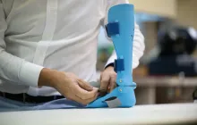
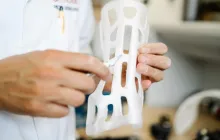
Material science lies at the heart of the ability to produce meaningful cardiac models. The heart’s tissues have a spectrum of mechanical properties, from the stiff, calcified annulus to the compliant myocardium and the delicate, pliable valve leaflets. Replicating this variety requires a careful pairing of printing technologies with materials that can mimic both geometry and biomechanics. Early 3D printed cardiac models relied on rigid plastics that provided crisp geometry but minimal physiological realism. As demand grew for models that could simulate tissue handling and device interactions, elastomeric polymers, flexible resins, and silicone-like materials emerged as viable options. These materials can be tuned to achieve specific shore hardness values and elongation behavior, enabling simulations that approximate the feel of cardiac tissue under manipulation. Multi material printers further broaden the possibilities by enabling the combination of stiff and compliant regions within a single model, which is particularly useful for reproducing interfaces between calcified deposits and healthier tissue or for delineating the boundary between a prosthetic ring and native myocardium. The choice of material also extends to transparency, which can be critical for visualizing internal pathways, channels, or the space where a device will be deployed. Transparent components allow clinicians to observe flow patterns, guidewire trajectories, and the interaction of devices with the surrounding anatomy in a way that is not possible with opaque models. Beyond passive geometry, researchers are exploring the incorporation of dynamic components, such as actuated elements or fluid circuits, that can simulate cardiac motion, ventricular filling, and pressure changes during the cardiac cycle. While achieving such functional realism presents technical challenges, advances in soft robotics, hydrogel composites, and embedded sensors hold promise for creating interactive, high-fidelity simulations that closely approximate in vivo conditions. The evolution of materials also intersects with sterilization considerations, biocompatibility requirements, and regulatory pathways, since some materials must withstand clinical use or experimental testing without compromising safety or performance. In practice, material selection is guided by the intended use case, balancing the need for mechanical realism, visualization clarity, cost, and manufacturability within the clinical workflow. As the field progresses, ongoing collaborations between material scientists, engineers, and clinicians will be essential to push the boundaries of what is possible while maintaining a pragmatic approach to adoption and reimbursement.
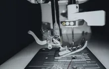
Printing technologies span a range of approaches, including vat photopolymerization, fused deposition modeling, selective laser sintering, and extrusion based systems. Each method has its own advantages in terms of resolution, speed, surface finish, and material compatibility. For heart models requiring tens of micrometers of detail, vat polymerization can deliver fine features that accurately reproduce valve leaflets and small vascular channels. For durable, reusable training aids, fused deposition modeling offers robust performance with a broader selection of polymers. In cases where flexible, elastic behavior is indispensable, soft elastomeric printing or silicone casting may be employed to capture the compliant nature of myocardium and vessel walls. The decision between a single poly material or a multi material, voxel-based printing strategy depends on the clinical objective, the desired biomechanical fidelity, and the facility’s technological maturity. Post processing, including cleaning, curing, and optionally mechanical finishing, further shapes the final product and can influence dimensional tolerance. The ongoing development of faster printing workflows, higher material libraries, and more predictable shrinkage and warping characteristics will continue to reduce production times and improve the reliability of the produced models. As printers become more accessible and user friendly, the integration of in-house printing capabilities within cardiology divisions is likely to grow, enabling more rapid iteration, customization, and experimentation in both clinical and educational settings.
In the realm of research and development, novel printing strategies are being explored to push 3D printing closer to living tissue. Bioprinting, which uses cell-laden bioinks to create tissue-like constructs, is an area of intense investigation with potential implications for modeling myocardial tissue, vascular networks, and scar formation. While these developments are still largely experimental and subject to stringent regulatory oversight, they highlight a trajectory toward increasingly realistic models that can be used not only for planning but also for studying pathophysiology and testing therapies in a controlled environment. For now, the majority of daily clinical use remains focused on high-fidelity structural models created from inert materials that accurately represent geometry and mechanical stiffness without engaging living cells. This pragmatic approach balances feasibility, safety, and clinical value while providing a stable platform for routine practice. The field will benefit as researchers continue to optimize materials for more authentic biomechanical behavior and as printing technologies mature to deliver faster, more consistent results with lower costs, broadening access to diverse patient populations and clinical settings.
Regulatory, Ethical, and Economic Considerations
As 3D printing becomes more integrated into cardiovascular care, regulatory and ethical considerations gain prominence. Regulatory bodies emphasize the need for quality management systems that address data integrity, model accuracy, material safety, and traceability. Institutions that produce patient-specific models must implement robust documentation of the imaging inputs, segmentation decisions, model specifications, and printing parameters to support clinical decision making and potential audits. Clear delineation of responsibility is essential, particularly when models influence treatment choices or device selection. In many jurisdictions, the responsibility for the clinical interpretation of a model rests with the treating physician rather than the designer of the model, but collaborative oversight ensures that the model remains a reliable surrogate for decision support rather than an autonomous clinical input. Ethical considerations extend to patient consent for the use of personal imaging data in generating physical replicas, with explicit discussion of potential benefits, limitations, and privacy protections. Economic considerations are equally important, as the value proposition for 3D printing depends on a balance between upfront costs, per-model expenses, and the downstream savings from improved planning, shorter procedure times, reduced complications, and enhanced education. Institutions must evaluate whether the investment supports measurable improvements in patient outcomes, workflow efficiency, and training capabilities. Health systems increasingly seek cost-effectiveness analyses that quantify the return on investment, taking into account not only direct monetary savings but also intangible benefits such as patient satisfaction and clinician confidence. Reimbursement frameworks are evolving, and advocates argue that, for selected indications and appropriate workflows, 3D printed models should be recognized as a reimbursable educational or planning tool similar to other imaging-based simulations. The sustainable integration of this technology will require thoughtful governance, ongoing data collection, and collaboration among clinicians, administrators, payers, and regulatory authorities to ensure that adoption is purposeful, scalable, and aligned with high standards of patient safety and care quality.
Quality assurance processes are critical to maintaining consistency across cases. Institutions often implement internal checklists that accompany the printing workflow, including verification steps for imaging source quality, segmentation accuracy, and dimensional fidelity. Periodic audits help identify sources of error, such as segmentation leakage or printer calibration drift, and guide corrective actions. Standardizing terminology and metrics allows for cross-institution comparisons and multi center research that can strengthen the evidence base for 3D printing in cardiology. Research initiatives increasingly emphasize patient outcomes, cost analyses, and educational impact as core endpoints, seeking to quantify how much value a model adds in specific clinical scenarios. The evolving regulatory landscape will likely introduce more formalized pathways for approving and integrating 3D printed models into care protocols, with particular attention to safety, effectiveness, and patient privacy. As this field matures, the alignment of clinical needs, technological capabilities, and policy frameworks will determine how broadly 3D printing is adopted and sustained across diverse cardiovascular programs.
Case Studies and Real-World Impact
Several compelling case studies illustrate how patient-specific 3D printed cardiac models have influenced real-world decisions. In a complex catheterbased procedure involving a heavily calcified aortic valve with an unusual annular geometry, a preoperative model allowed the team to rehearse the deployment and select a device size and approach with greater confidence, leading to a smoother intraoperative course and a shorter fluoroscopy time. In a pediatric patient with a rare combination of congenital defects, a life-sized heart replica served as a central element in planning staged repairs, enabling a more precise sequence of interventions and a clearer understanding among the multidisciplinary team about potential interactions between the defects. In adult congenital heart disease, models that combined anatomy with scalable flow channels supported simulations of coronary perfusion and helped identify risks related to bypass strategies, contributing to a more individualized plan that balanced risk and benefit. Training programs have also benefited from case-based simulations, where learners can work through the steps of a repair or a transcatheter intervention with a tactile reference that anchors theoretical concepts to concrete anatomy. Across these experiences, the consistent theme is that 3D printed models complement traditional imaging by providing a hands-on, patient-specific reference that can reduce uncertainty, facilitate collaboration, and enhance communication with patients and families. While not every case requires a printed model, there is growing recognition that, for certain anatomies, therapies, or educational goals, a well crafted model can meaningfully tilt the balance toward safer, more precise care and improved outcomes. The growing body of real-world evidence supports a nuanced approach, where the decision to print is guided by clinical complexity, anticipated benefit, and resource considerations rather than a one-size-fits-all expectation.
Beyond individual cases, health systems are documenting aggregated data on the impact of 3D printing in cardiology. Early experiences highlight reductions in procedure times and radiation exposure when complex anatomies are anticipated with the help of models, along with improved patient comprehension and satisfaction. Some programs report that printing enables better coordination during multidisciplinary conferences, where different specialties review anatomy from multiple perspectives and align on an agreed plan. Others emphasize the educational value for trainees as a catalyst for more thorough preoperative analysis, which may translate into improved procedural planning and safer execution. While the literature continues to mature, the practical messages from these real-world experiences are clear: 3D printing in cardiology is not a universal remedy, but when applied to appropriately tailored questions, it can yield tangible benefits across planning, education, and patient engagement. As centers accumulate experience and share outcomes, best practices will emerge, and the technology will become more efficiently integrated into standard cardiovascular care pathways. This iterative learning process promises to refine patient selection, optimize workflows, and ensure that the technology remains aligned with the overarching goals of safety, efficacy, and compassionate, patient-centered care in cardiology.
Future Directions and Innovations in Cardiac 3D Printing
The horizon for 3D printing in cardiology is bright with avenues for refinement and expansion. One area of ongoing development is the push toward more realistic biomechanical properties, where materials and printing strategies enable models to mimic the non linear elastic behavior of cardiac tissues. Researchers are exploring dynamic models that can emulate pulsatile flow, chamber motion, and valve dynamics to create reproducible laboratory environments for device testing and procedural rehearsal. The integration of printed models with computational simulations can create hybrid platforms where anatomical fidelity is coupled with fluid-structure interaction analyses, enriching our understanding of how devices interact with the heart under physiologic conditions. Such hybrid approaches may enable more precise predictions of hemodynamic changes following interventions, guiding decision making with a combination of tangible and computational evidence. In addition to mechanical realism, advances in imaging co registration, color coding, and labeling within models will help clinicians identify critical structures quickly, improving workflow efficiency and reducing cognitive load during high-stakes planning sessions. The emergence of more compact, cost-effective printers with broader material catalogs will democratize access to 3D printing, extending its reach to community hospitals and smaller centers that currently face barriers to adoption. This expansion will support equity by enabling more patients to benefit from patient-specific planning even when they are geographically distant from major specialized centers. Educationally, the continued expansion of curricula and training pathways will prepare the next generation of cardiologists not only to interpret complex imaging but to leverage 3D printing as an integral component of diagnostic reasoning and procedural mastery. The confluence of print technology with sensorized materials, soft robotics, and tissue-mimicking composites holds the promise of increasingly realistic simulators that can transform preclinical testing and therapeutic development into safer, more effective, and more efficient processes. As these innovations unfold, careful attention to patient safety, ethical use, and cost management will ensure that the benefits of 3D printing are realized in ways that are sustainable and broadly beneficial across diverse patient populations. The role of policy makers, payers, researchers, and clinicians will be pivotal in shaping how quickly and how deeply these technologies become embedded in routine cardiology practice, with patient welfare as the enduring compass guiding every advancement.