The landscape of organ transplantation has long been characterized by life changing successes tempered with persistent shortages and complex ethical questions. For decades, patients facing organ failure have depended on donor availability, waiting lists, and the delicate balance between urgency and quality of life. In this context, three dimensional bioprinting has emerged as a field promising to bridge the gap between rising demand and the supply of functional, compatible tissues and organs. The concept encompasses the precise placement of living cells, supportive biomaterials, and signaling molecules in a manner that mimics the architecture of native tissues. This convergence of biology and engineering holds the potential to shift the paradigm of transplantation from a dependency on cadaveric or animal sources toward patient specific, lab generated grafts, or at least functional components that can restore organ function while reducing immunological barriers. The prospects of bioprinted organs extend beyond replacement; they include models for drug testing, disease modeling, and regenerative strategies that could ultimately lessen the burden on healthcare systems and extend lives in ways that were unimaginable even a decade ago. The promise is tempered by the need to overcome formidable scientific, clinical, and regulatory challenges, but the trajectory of ongoing research suggests that bioprinting will increasingly inform, augment, and perhaps redefine the process of organ transplantation in the years ahead.
Historical context and the evolution of bioprinting concepts
The journey toward bioprinting as a practical tool for organ replacement can be traced through the broader evolution of additive manufacturing. Early three dimensional printing emerged as a rapid prototyping method that enabled designers to translate complex forms into tangible objects with speed and reproducibility. As scientists recognized the possibility of incorporating living cells and biologically active substances into printed constructs, a new field known as bioprinting took shape. The conceptual leap was to move from inert plastic parts to biologically functional tissues that could integrate with and respond to a living organism. Over time, researchers developed and refined a set of printing modalities, including extrusion based systems that lay down viscous bioinks containing cells, inkjet style printers that deposit droplets with precision, and laser assisted approaches that enable high resolution placement of delicate cell assemblies. Each modality offered distinct advantages and faced particular limitations in terms of cell viability, structural complexity, and scalability. This historical arc is marked by incremental demonstrations of tissue primitives such as thin sheets of living cells, vascular networks in simplified channels, and small tissue constructs that behaved in a patient relevant manner under laboratory conditions. The cumulative knowledge from these early steps set the stage for attempts to print organ level structures that would require a combination of precise geometry, robust biomaterials, and viable, functional cell populations capable of maintaining homeostasis in a transplanted environment. As the field matured, attention turned to the aspects of integration with host physiology, immunological compatibility, and the dynamic demands of perfusion, mechanical load, and long term viability. The historical narrative therefore blends engineering ingenuity with cell biology, materials science, and a careful regard for clinical realities, illustrating how a multidisciplinary effort can transform a theoretical possibility into a practical research program with tangible implications for organ transplantation.
The core technologies behind bioprinting and how they apply to organs
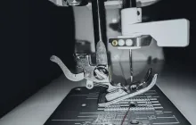
Bioprinting relies on a suite of technologies designed to assemble living tissues with controlled architecture. In extrusion based bioprinting, cells suspended in a hydrogel like bioink are extruded through a nozzle to form filaments that assemble into three dimensional constructs. This approach emphasizes scalability and the ability to handle relatively high cell densities, making it well suited for larger tissue regions that may require substantial mechanical support. Inkjet based bioprinting, by contrast, dispenses discrete droplets of bioink with high temporal resolution, enabling rapid patterning and fine spatial control, though it often requires optimization to preserve cell viability at the nozzle and during deposition. Laser assisted bioprinting uses a focused laser to propel cellular material from a donor substrate onto a receiving surface, offering high resolution and gentler handling for sensitive cell types, but demanding precise calibration to avoid thermal or mechanical damage. Each modality has implications for organ scale and functionality: extrusion can build voluminous structures with viscoelastic materials, inkjet can achieve detailed cellular patterning at a micro scale, and laser assisted approaches can support the placement of multiple cell types with spatial fidelity. Beyond the printing hardware, the materials science that underpins bioinks is equally critical: bioinks must provide a hospitable environment for cell viability and proliferation, permit nutrient and waste exchange, and progressively remodel in concert with host tissues. The synergy of printer design, bioink chemistry, and biophysical cues shapes the mechanical properties and long term behavior of printed grafts, from the elasticity of a soft organ matrix to the stiffness required for load bearing portions of a transplanted organ.
Bioinks, cells, and the construction of vascularized tissue
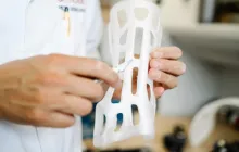
At the heart of any successful bioprinted organ is the careful orchestration of cells and biomaterials to recreate tissue specific microenvironments. Bioinks are composed of biopolymers that provide a scaffold like matrix, while sustaining cells with a hospitable chemical milieu. For organ engineering, bioinks must accommodate a dynamic range of requirements: viability during printing, appropriate mechanical cues, and the capability to support differentiation and maturation toward tissue specific phenotypes. In transplants, the inclusion of patient derived cells holds particular appeal because it can reduce the risk of rejection and immunological complications. The patient specificity of cells, when feasible, integrates with personalized medicine strategies that seek to tailor grafts not only to anatomical size and shape but to the unique cellular and genetic makeup of the recipient. Creating a viable organ requires more than embedding cells within a static scaffold; it demands a functional vasculature that can deliver oxygen and nutrients to thousands or millions of cells while removing waste. Early proofs of concept demonstrated perfusable networks within printed tissues, but scaling these networks to organ level sizes and achieving stable perfusion over months and years remains a central challenge. The emergence of angiogenic factors, microfluidic channel design, and decellularized extracellular matrices as supporting frameworks has advanced the potential of bioprinted organs, offering pathways to integrate native blood vessel formation with printed constructs and to align mechanical properties with the demands of organ physiology.
Vascularization and perfusion: the bottleneck toward whole organ printing
One of the most persistent bottlenecks in translating bioprinted tissues to transplantable organs is the establishment of a functional vascular system. In native organs, blood vessels permeate every region of tissue, enabling rapid delivery of oxygen and nutrients and facilitating waste removal. In a printed construct, diffusion limits rapidly constrain cell survival beyond a certain thickness, creating a need for perfusion networks that mimic microcirculation. Various strategies have emerged to address this. Some researchers embed sacrificial materials within printed structures that can be removed after printing to yield interconnected channels that can be perfused with nutrient solutions. Others co print endothelial cells arranged along network like geometries with pre patterned lumens and connect them to external perfusion circuits. Still other approaches leverage decellularized organ matrices that retain a native vascular architecture, providing a realistic scaffold that can guide repopulating cells while preserving channels for perfusion. The ultimate goal is to achieve a perfusion compatible architecture that remains patent and functional after implantation. Realizing this in a whole organ, with its complex hierarchy of arteries, arterioles, capillaries, and venules, requires integrating biophysical cues, immune tolerance, and physiological feedback mechanisms to sustain viability under the mechanical and metabolic loads of the living body. Success in vascularization would enable longer term graft survival and open doors to printing larger, more sophisticated organs such as kidneys, livers, or hearts, where perfusion is essential to function and longevity.
Material science, immunology, and compatibility considerations
The interaction between a bioprinted graft and the host immune system is a central determinant of clinical success. Materials used in bioinks range from naturally derived polymers to synthetic polymers, each with distinct degradation profiles, mechanical properties, and bioactive signals. The ideal bioink would be biocompatible, non immunogenic, and capable of gradually transferring biological control from the printed scaffold to the host tissue as remodeling occurs. In addition to the biophysical compatibility, the immunological perspective in transplantation extends to the source of cells: autologous cells derived from the patient are theoretically ideal for minimizing rejection, yet challenges in expanding sufficient numbers of patient specific cells and driving them toward desired organ specific lineages persist. When allogeneic or induced pluripotent stem cell based approaches are used, strategies to reduce immunogenicity become essential, including immune cloaking of grafts, selective immune modulation, and careful design of extracellular matrices to present non inflammatory cues. The materials science dimension also encompasses mechanical stability and long term durability; printed constructs must endure physiological strains, dynamic fluid flow, and chemical environments within the body, all while gradually integrating with native tissues. Regulatory considerations intersect with these scientific realities, necessitating rigorous evaluation of material safety, degradation byproducts, and potential immune responses over extended timeframes. The synergy of immunology and materials science thus defines a critical corridor toward clinically meaningful bioprinted organs.
Mechanical properties and organ specific challenges
Organs possess distinctive mechanical signatures that reflect their function and location. A heart graft experiences rhythmic pulsatile loads, while a liver endures shear stress and compressive forces within the abdominal cavity, and a kidney processes fluctuating fluid pressures. Matching these mechanical environments in a printed construct is essential to achieve proper function and durability. To approach this, researchers tailor hydrogel stiffness, viscoelastic behavior, and porosity to mimic native tissue. They also consider the anisotropy of tissues, where properties vary along different directions due to fiber orientation and cellular alignment. In cardiac tissue, for instance, contractile alignment of cardiomyocytes and the formation of gap junctions are critical for synchronous beating, while in kidney tissue the architecture of nephrons and tubules demands precise patterning and robust filtration interfaces. The complexity grows when combining multiple tissue types into a single organ system, prompting multi material printing approaches and modular strategies where smaller functional units are assembled into an integrated whole. The interplay between mechanical cues and cellular responses shapes maturation, function, and ultimately the success of an organ transplant scenario. Addressing these mechanical challenges requires iterative engineering, advanced imaging, and computational modeling to predict how printed constructs will behave in vivo and how they will adapt post implantation.
Bioreactors, conditioning, and maturation of printed grafts
Biological maturation of printed tissues often depends on exposure to controlled biological environments that supply nutrients, apply mechanical stimuli, and recreate physiologic signals outside the body before implantation. Bioreactors designed for this purpose can deliver perfusion, oxygenation, and cyclic stretch or shear, promoting tissue organization and functional maturation. Conditioning protocols aim to transition a printed scaffold from a laboratory state to a more physiologically relevant state, approaching the performance of a native organ. This pre conditioning may include electrical stimulation to encourage cardiomyocyte alignment and function, or pulsatile flow to condition vascular networks. The goal is to produce grafts that exhibit stable structure, persistent viability, and measurable functional output prior to transplantation. Beyond technical merit, bioreactor conditioning also contributes to quality control, enabling assessment of mechanical integrity and biological activity under controlled conditions. The maturation process remains an active research area, with ongoing investigations into the optimal timing, stimuli, and culture environments required to render a bioprinted organ ready for the dynamic, demanding setting of the human body.
Clinical translation: current landscape and near term prospects
The translation of bioprinting from the laboratory bench to the clinical bedside is a carefully staged journey that navigates scientific validation, manufacturing scalability, patient safety, and regulatory scrutiny. At present, the most tangible achievements involve tissue constructs and organ analogs used primarily for research, drug testing, and preliminary translational studies rather than fully transplantable organs. Nonetheless, incremental progress in fabricating vascularized tissue patches and organ segments demonstrates the feasibility of certain concepts in living systems and provides valuable insights into designing more complex grafts. The near term prospects emphasize refining printing techniques, standardizing bioinks, optimizing cell sourcing, and validating functional endpoints relevant to specific organ applications. Clinically viable bioprinted grafts may first appear as partial organs, such as sections of liver tissue or perfusable vascular networks that could support existing organ function or serve as ready to transplant components in combination with conventional organs. The path to a fully printed organ that can replace an entire native organ without immunosuppression would likely require sustained breakthroughs in vascular integration, scalable manufacturing, long term durability, and regulatory alignment. While cautious optimism remains warranted, the trajectory of ongoing research points toward a future in which bioprinted elements contribute to transplantation in meaningful, complementary ways well within the next decade.
Ethics, regulation, and safety considerations in bioprinted organs
Bioprinting raises a spectrum of ethical questions that accompany any transformative medical technology. Issues include ensuring equitable access to advanced therapies, protecting patient autonomy, and managing expectations about what bioprinted organs can achieve. Safety considerations are equally paramount, with thorough preclinical testing required to evaluate tumorigenicity, unintended differentiation, inflammatory responses, and the risk of infection. Regulatory frameworks increasingly recognize the complexity of bioprinted products, often treating them as combination products that involve cells, biomaterials, and devices. This necessitates a cross disciplinary assessment to address quality control, standardization of manufacturing practices, and post market surveillance. The ethical and regulatory discourse thus centers on balancing innovation with patient protection, transparency about capabilities and limitations, and the creation of pathways that encourage responsible development while preserving patient safety as a foundational priority. Collaboration among scientists, clinicians, policymakers, and patient advocates is essential to translating the promise of bioprinting into therapies that are both effective and ethically sound.
Applications beyond whole organs: niches, patches, and disease models
Even before the achievement of fully functional printed organs, bioprinting has found valuable applications in creating tissue patches that can support organ function or repair damaged areas. Cardiac patches designed to deliver electrical and mechanical stimulus, liver tissue approximations that assist metabolic processes, and pancreatic islet arrays used for diabetes research exemplify how printed constructs can contribute to patient care. In addition, bioprinted tissues serve as advanced disease models that enable researchers to study pathophysiology and test therapeutics in a human relevant context, reducing reliance on animal models and accelerating drug discovery. These intermediate applications not only provide direct clinical benefits when appropriate but also act as stepping stones that refine printing strategies, bioink formulations, and maturation protocols necessary for eventual organ level applications. The synergy between regenerative medicine, pharmacology, and organ transplantation becomes increasingly evident as bioprinting moves through successive layers of complexity and clinical relevance.
Interdisciplinary collaboration and the emergence of new centers of expertise
The push toward clinically meaningful bioprinted organs necessitates a convergence of disciplines that traditionally function in separate silos. Tissue engineering, cellular biology, materials science, mechanical and chemical engineering, computer assisted design, and clinical medicine must collaborate seamlessly to design, print, test, and evaluate complex grafts. This collaborative ecosystem fosters the development of specialized training programs, shared research facilities, and cross disciplinary funding mechanisms that accelerate progress while maintaining rigorous scientific and ethical standards. As institutions invest in multidisciplinary laboratories and clinical trial pathways, the culture of innovation shifts toward a holistic view of transplantation that integrates printing as a core tool rather than a distant, experimental concept. The emergence of consortia and public private partnerships further catalyzes the translation process, enabling larger data sets, standardized protocols, and more robust safety assessments that are essential to regulatory approval and patient acceptance.
Patient impact, accessibility, and the societal horizon
The potential impact of 3D bioprinting on patient lives extends beyond the elimination of waiting lists. By enabling personalized grafts that align with individual anatomy and cellular makeup, bioprinting can offer improvements in graft survival, reduced need for lifelong immunosuppression, and better overall quality of life. However, the realization of these benefits hinges on balancing cost and access, ensuring that advanced therapies do not become the exclusive domain of affluent sectors. Healthcare systems will need to adapt to new production pipelines, regulatory requirements, and ethical considerations. Training clinicians to interpret and manage bioprinted grafts, updating infrastructure to support biomanufacturing, and establishing reimbursement frameworks that reflect the distinct value proposition of printed organs will be critical steps in broadening access. As research progresses, public engagement and transparent communication about realistic timelines, potential risks, and expected outcomes will be essential to maintaining trust and guiding policy decisions that shape the equitable deployment of this technology.
Future directions: what must be achieved to make printing whole organs routine
Charting the future of bioprinting in organ transplantation involves identifying and pursuing several key milestones. Achieving robust and durable vascular networks that can support organ metabolism over months and years is paramount. Advancements in bioink chemistry, including smart materials capable of responding to biochemical cues and adapting stiffness in response to remodeling, may enhance integration with host tissues. Developments in stem cell biology and directed differentiation can provide sources of organ specific cells that avoid ethical and immune concerns while maintaining genomic stability. Computational modeling and machine learning will play increasing roles in optimizing print paths, predicting maturation trajectories, and forecasting graft performance under physiological conditions. Moreover, regulatory science must evolve to accommodate the complex nature of bioprinted products, incorporating rigorous preclinical validation, clear definitions of quality attributes, and streamlined, patient centered pathways for clinical testing. The collaborative ecosystem will need to align educational programs, manufacturing capabilities, and clinical trial infrastructure to transform theoretical potential into safe, accessible, and effective organ replacement solutions. The envisioned horizon includes not only fully functional transplanted organs but also hybrid approaches that combine printed tissue components with conventional organ platforms to extend function and longevity, ultimately reshaping the standard of care for organ failure across diverse patient populations.
Safety, quality control, and manufacturing considerations at scale
As bioprinting moves closer to widespread clinical use, the imperative to ensure safety and consistent quality becomes more pressing. Quality control frameworks for bioprinted organs will need to address cell viability pre and post printing, the uniformity of tissue architecture, the integrity of vascular networks, and the long term stability of implanted grafts. Manufacturing scale introduces batch to batch variability that must be minimized through standardized bioink formulations, precise printer calibration, and robust environmental controls. Sterile production environments, validated sterilization methods, and traceable supply chains will underpin patient safety. Additionally, rapid, nondestructive evaluation techniques that can assess functional attributes of printed tissues before implantation will help reduce risk and support regulatory submissions. The integration of quality management systems with clinical workflows will be essential to maintain high levels of safety, efficacy, and reproducibility as bioprinted organs transition from research settings to hospital use.
Closing perspective: the evolving role of bioprinting in organ transplantation
In summary, 3D bioprinting represents a transformative approach to organ transplantation that complements traditional donor based strategies with design driven, patient centered, and potentially immunomodulated grafts. While the field faces substantial scientific and regulatory hurdles, the progress achieved across modalities, bioinks, vascularization strategies, maturation protocols, and translational frameworks demonstrates a clear and steadily widening path toward clinically meaningful outcomes. The importance of continued collaboration across disciplines, investment in infrastructure and training, and thoughtful engagement with ethical and policy questions cannot be overstated. The journey toward routine bioprinted organs will be gradual and iterative, marked by incremental breakthroughs and careful demonstrations of safety, function, and patient benefit. As researchers, clinicians, and policymakers navigate this evolving landscape, the central promise remains steadfast: to offer new hope for people facing organ failure by harnessing the power of three dimensional bioprinting to create living, functional tissue that can harmonize with the human body and extend the possibilities of what transplantation can achieve.