Medical imaging has long stood at the intersection of science, engineering, and clinical care, continually transforming how doctors visualize the human body, identify disease, and guide therapy. The journey began with simple projections that revealed silhouettes of internal structures and gradually evolved into sophisticated systems capable of revealing physiology, metabolism, and molecular processes with astonishing precision. Each era brought new detectors, new computational approaches, and new physics that broadened the scope of what could be observed noninvasively. The current landscape is a tapestry woven from breakthroughs in hardware design, signal processing, data analytics, and interdisciplinary collaboration among radiologists, physicists, engineers, and informaticians. This ongoing evolution has not only improved diagnostic accuracy but also reshaped patient pathways, enabling earlier detection, safer imaging, and more effective, individualized treatment strategies across a wide range of diseases.
From the earliest radiographs to the advent of magnetic resonance imaging and beyond, imaging technologies have consistently pursued two parallel goals: to enhance visibility of relevant tissues and to minimize risk to patients and healthcare workers. In the early chapters of medical imaging, the discovery of X rays in the late nineteenth century opened a window into the anatomy that could be explored with relative ease, quickly proving invaluable in diagnosis and management. As computing power expanded, computed tomography emerged, producing cross sectional views that could be reconstructed into three dimensional representations, thereby enabling clinicians to interrogate complex anatomy with unprecedented clarity. Later, the competition between different physical principles led to the rise of magnetic resonance imaging, which exploited the behavior of hydrogen nuclei in a magnetic field to generate high contrast images of soft tissues without ionizing radiation. The cumulative effect of these milestones was to shift imaging from a static snapshot to a dynamic, multi dimensional description of structure and function, laying the groundwork for a more precise, quantitative, and repeatable form of medical visualization that would become central to modern clinical practice.
Multimodal imaging and the rise of integrated diagnostics
One of the most transformative trends in medical imaging has been the integration of complementary modalities into cohesive diagnostic workflows. The combination of molecular information with high spatial resolution, achieved through hybrid systems such as PET/CT, fused metabolic data with anatomical context, and allowed clinicians to localize biologic processes within precise structural landmarks. The introduction of PET/MRI added a further dimension by uniting metabolic insight with superior soft tissue contrast and functional imaging capabilities, thereby enabling more accurate characterizations of certain tumors, inflammatory processes, and neurodegenerative conditions. This multimodal framework extends beyond mere co-registration; it fosters a unified interpretive paradigm in which quantitative metrics, spatial correlations, and temporal dynamics are synthesized to inform prognosis, treatment planning, and response assessment in a way that neither modality could achieve alone.
As imaging technology matured, the emphasis shifted toward developing integrated diagnostic ecosystems that streamline acquisition, processing, and interpretation. Advanced image fusion algorithms, standardized protocols for cross modality calibration, and cross-disciplinary interpretation teams have become essential features of contemporary radiology departments. The clinical impact is evident in improved lesion conspicuity, more accurate staging for cancers, and enhanced monitoring of therapeutic efficacy. In addition, multimodal imaging has begun to empower guidance for interventions and therapies, where precise localization of target tissue and real-time feedback on treatment effects can influence procedural decisions. The net effect of this convergence is a more holistic view of disease biology, where anatomy, physiology, metabolism, and molecular markers are brought together within a single diagnostic narrative that informs patient-specific care plans.
Advances in detector technology and image acquisition
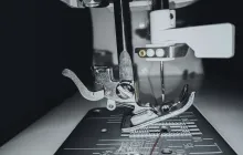
Progress in detector science has been a driving force behind sharper images, shorter scan times, and lower radiation exposure. The shift from traditional film to digital detectors, followed by flat-panel detectors, dramatically improved sensitivity, dynamic range, and the ability to rapidly capture high spatial resolution data. Contemporary detectors employ solid-state technologies and sophisticated materials to optimize photon collection efficiency, energy discrimination, and timing accuracy, enabling not only crisper anatomical imaging but also specialized capabilities such as spectral separation and energy-resolved imaging. In parallel, improvements in gantry design, patient positioning, and motion correction strategies have enhanced robustness to patient motion and physiological variability, expanding the range of patients who can benefit from advanced imaging, including children and those with limited cooperation.
Another pivotal advance is the emergence of photon-counting detectors, which promise to transform computed tomography by directly counting individual photons rather than integrating energy across a detector element. This capability improves image quality, reduces noise, and provides material-specific information that can support tissue characterization and dose optimization. The impact extends to increasingly sophisticated spectral imaging, where multiple energy thresholds enable the separation of tissues and contrast agents, enabling clinicians to distinguish between overlapping structures with greater confidence. As these detectors move from research laboratories into routine clinical use, they are poised to redefine how clinicians acquire, interpret, and apply CT data across a broad spectrum of indications, from emergency imaging to oncologic assessment and beyond.
Functional and molecular imaging technologies
A key theme in modern imaging is the shift from purely anatomical depictions toward functional and molecular descriptions of tissue biology. Functional MRI, which measures blood oxygen level dependent signals, has become a cornerstone in neuroscience research and clinical evaluation of brain pathology, enabling inferences about neural activity and connectivity that correlate with cognitive function and disease states. Diffusion-weighted imaging and diffusion tensor imaging extend this capability by probing microstructural properties of tissue, revealing pathways and integrity in white matter tracts and offering insights into acute injuries, tumor infiltration, and developmental changes. On the molecular side, positron emission tomography and its array of radiotracers illuminate metabolic processes and receptor expression, providing a window into tumor biology, infection, and neurodegeneration that is complementary to structural imaging.
The convergence of functional, structural, and molecular data supports a higher level of diagnostic precision and a deeper understanding of disease mechanisms. Radiomics and quantitative imaging biomarkers extract meaningful patterns from complex datasets, enabling more objective assessments of tumor aggressiveness, treatment response, and prognosis. In oncology, for example, dynamic contrast enhanced imaging tracks perfusion and permeability, while diffusion metrics reflect cellular density and membrane integrity, offering surrogate endpoints for early therapeutic efficacy. In neuroimaging, functional and metabolic insights contribute to distinguishing atypical presentations, guiding surgical planning, and monitoring longitudinal changes that correlate with clinical outcomes. The ongoing refinement of acquisition protocols, data processing pipelines, and validation studies is essential to translate these rich datasets into routine clinical decision support.
Ultrasound innovations and real-time guidance
Ultrasound imaging remains a versatile workhorse due to its portability, safety profile, and real-time capabilities. Recent advances have expanded its role from conventional structural assessment to dynamic assessment, functional evaluation, and image-guided therapy. Three-dimensional and four-dimensional ultrasound provide volumetric visualization and real-time motion tracking that enhance fetal imaging, obstetric assessments, and guidance for procedures such as biopsies and injections. Doppler techniques continue to evolve, offering more sensitive assessments of blood flow and hemodynamics in vascular and cardiac conditions, while elastography adds a tissue stiffness dimension that improves lesion characterization and risk stratification.
Portable and pocket-sized ultrasound devices have made point-of-care imaging a routine part of clinical workflows, enabling rapid bedside assessment in critical care, emergency medicine, and rural or resource-limited settings. These devices increasingly integrate artificial intelligence through automated image optimization, artifact suppression, and preliminary interpretation aids, which can reduce operator dependency and support non-specialist clinicians in obtaining useful information. In interventional contexts, fusion of ultrasound imaging with other modalities or navigation systems enhances precision during procedures, improving safety and outcomes. The cumulative effect of these ultrasound-focused advancements is a broader, more accessible imaging toolkit that complements larger modalities and accelerates clinical decision-making in diverse environments.
Magnetic resonance imaging innovations
Magnetic resonance imaging continues to push the boundaries of spatial and contrast resolution while expanding functional and metabolic assessment. High-field and ultra-high-field systems, including 3T and 7T platforms, offer improved signal-to-noise ratios that unlock finer anatomical detail and more sensitive functional measurements, though they also present challenges related to susceptibility effects and patient comfort. Advances in fast imaging sequences reduce scan times, minimize motion artifacts, and enable dynamic studies such as perfusion imaging, spectroscopy, and perfusion maps of organs beyond the brain. Diffusion weighted imaging and tractography remain central to neurological and oncologic applications, where microstructural insights help delineate tumor margins, monitor treatment response, and chart disease progression with greater confidence.
MR fingerprinting is a notable example of quantitative imaging that derives multiple tissue properties from a single, efficient acquisition, providing robust T1 and T2 maps, proton density, and other parameters that facilitate standardized tissue characterization. Parallel transmission and received array coils improve uniformity and depth penetration, broadening the scope of high-quality imaging to regions previously challenging to image in detail. Functional MRI and spectroscopy extend MRI's reach into functional biology, enabling studies of brain connectivity, metabolic processes, and tissue pathology that enrich clinical interpretation and research investigations. Together, these innovations elevate MRI from a primarily anatomical modality to a comprehensive platform for assessing structure, function, and metabolism in health and disease.
Computed tomography breakthroughs and dose-conscious design
Computed tomography has undergone a renaissance driven by efforts to balance diagnostic richness with patient safety. Iterative reconstruction algorithms reduce noise and artifacts at low radiation doses, while model-based approaches improve image fidelity in challenging regions such as the sinuses or the chest. The emergence of spectral or dual-energy CT allows simultaneous acquisition at multiple energy spectra, enabling material discrimination and precise characterization of tissues and contrast agents. This capability enhances lesion detection, improves contrast between abnormal and normal tissue, and supports tasks ranging from kidney stone characterization to cardiothoracic assessment and oncologic staging.
Three-dimensional CT angiography and high-resolution chest imaging have expanded the utility of CT in vascular and pulmonary diseases, while dose modulation strategies and careful protocol optimization minimize exposure during routine scans. The development of photon-counting detectors holds promise for further dose reductions and spectral separation, potentially enabling broader adoption of CT in sensitive populations such as pediatric patients. As CT technology matures, the clinical workflow benefits from faster acquisitions and better alignment with clinical questions, enabling clinicians to obtain high-quality information quickly, safely, and reproducibly in a variety of settings, from acute care to outpatient imaging centers.
Radiomics, AI in imaging interpretation, and clinical decision support
The rapid growth of artificial intelligence and machine learning has reshaped how imaging data are analyzed, interpreted, and translated into actionable clinical insights. Radiomics extracts high-dimensional features from images that may reflect tumor texture, heterogeneity, and microenvironment, providing quantitative biomarkers that can inform prognosis, therapy selection, and monitoring. Deep learning models support tasks such as lesion detection, segmentation, and classification, often achieving performance levels that complement expert interpretation and can accelerate workflow in high-volume centers. Importantly, these computational tools are increasingly integrated into validation pipelines, with emphasis on reproducibility, generalizability, and alignment with clinical endpoints.
Beyond interpretation, AI is increasingly embedded into image acquisition and reconstruction processes themselves. Intelligent systems can optimize parameter settings, correct motion, reduce artifacts, and predict the most informative sequences or views for a given patient, thereby increasing efficiency and reducing the burden on patients and clinicians. The clinical impact spans oncology, cardiology, neurology, and musculoskeletal imaging, where AI-augmented decision support is helping to standardize reporting, reduce variability, and enable earlier detection of diseases with subtle or heterogeneous presentation. As these tools become more pervasive, there is growing emphasis on transparency, explainability, and careful evaluation to ensure that AI augments rather than replaces the nuanced judgment of experienced clinicians.
Radiation safety, dose optimization, and patient-centric imaging
Historically, radiation exposure has been a central concern in imaging that uses ionizing radiation, driving a continuous push toward safer protocols without compromising diagnostic quality. Dose optimization approaches include automatic exposure control, smarter protocol selection, and patient-specific adjustments that tailor radiation dose to body habitus and clinical indication. The development of advanced reconstruction techniques and noise-reduction algorithms further enhances image quality at lower doses, reducing the cumulative radiation burden for patients who require repeated imaging, such as those undergoing cancer surveillance or congenital disease monitoring. Clinicians increasingly weigh the trade-offs between image detail, diagnostic confidence, and radiation risk, fostering a patient-centric culture that prioritizes safety alongside diagnostic accuracy.
Efforts to minimize radiation exposure intersect with improvements in alternative modalities and non-ionizing techniques, expanding the toolbox for clinicians while maintaining high standards of care. Ultrasound and MRI, which do not involve ionizing radiation, are increasingly used for follow-up imaging or initial screening in appropriate contexts, while CT protocols emphasize dose awareness during pediatric and elderly imaging. National and international guidelines continue to evolve, emphasizing standardized protocols, quality assurance, patient communication about risks and benefits, and the responsible adoption of new technologies that promise clinical gains without disproportionate safety concerns. In parallel, education and training emphasize best practices in shielding, dose tracking, and verification of equipment performance to sustain a culture of safety across imaging facilities.
Hybrid imaging platforms and image-guided therapy
The maturation of hybrid imaging platforms has expanded the clinical reach of imaging to therapeutic decision-making and image-guided interventions. PET/CT and PET/MRI provide metabolic context to precise anatomical localization, enabling clinicians to plan, monitor, and adapt treatments with a level of confidence not achievable with a single modality. In oncologic care, this synergy informs decisions about surgery, radiation therapy, and systemic treatments by revealing tumor biology, hypoxia, and proliferative activity in real time. The integration of imaging with therapeutic devices, including navigation-guided biopsy, targeted drug delivery, and image-guided radiotherapy, exemplifies how diagnostic tools can actively participate in treatment execution rather than serving solely as diagnostic endpoints.
Advances in real-time imaging during procedures, improved tracking algorithms, and augmented reality-assisted navigation are converging to make interventions safer and more precise. This trend is complemented by ongoing efforts to standardize interpretation across modalities, enabling clinicians to interpret dynamic imaging data in conjunction with static anatomic references. The clinical implications are far-reaching, allowing more accurate tumor delineation for surgery and radiotherapy, better assessment of margins, and timely evaluation of treatment response. As hybrid imaging ecosystems become more accessible and integrated into routine care, they hold the promise of enabling truly personalized, image-driven therapies that optimize outcomes while reducing unnecessary interventions and exposure to risk for patients.
Future directions and ethical considerations in imaging technology
Looking forward, the field of medical imaging is poised to embrace a more automated, data-intensive era while confronting important ethical and governance challenges. Advances in cloud-based analytics, federated learning, and secure data sharing promise to unlock collective knowledge from diverse patient populations, but they require careful attention to privacy, consent, and data ownership. Ensuring that AI and automation enhance equity in access to high-quality imaging across regions and populations will require deliberate policy, inclusive research designs, and transparent validation practices that address potential biases in training data or algorithmic outputs. Clinicians, researchers, and regulators will need to collaborate to establish robust standards for performance, safety, and accountability as new modalities, detectors, and computational tools reach clinical maturity.
Another facet of the evolving landscape involves patient engagement and education, including clear communication about radiation risks, the benefits of advanced imaging, and the expectations around test results. As imaging becomes increasingly data rich, clinicians must balance the thrill of discovery with the practical realities of clinical relevance, avoiding overdiagnosis and ensuring that imaging findings translate into meaningful improvements in patient outcomes. Interoperability and standards remain foundational, with initiatives aimed at harmonizing data formats, reporting schemas, and measurement scales so that insights generated in one institution can be validated and applied across the broader healthcare ecosystem. Finally, the expansion of educational programs for radiologists, technologists, and allied health professionals will be essential to sustaining innovation, ensuring safety, and maintaining the human-centered focus at the heart of medical imaging’s enduring mission.