Understanding kidney stones and how they form
Kidney stones are hard, crystalline mineral formations that develop inside the kidneys or urinary tract when certain substances in urine become concentrated enough to crystallize. They are not a single disease but a family of conditions that share the common feature of mineral buildup occurring in a fluid environment that cannot dissolve those minerals fully. The most common type is calcium-based stones, typically calcium oxalate, which arise when levels of calcium or oxalate are higher than normal or when urine becomes unusually acidic or concentrated. Other stones form from uric acid when urine is frequently acidic or when the body metabolizes purines excessively; struvite stones form in the presence of certain bacteria that raise the urine pH, and cystine stones occur in people with a genetic condition that causes high levels of certain amino acids in the urine. Each stone type has its own set of risk factors, but dehydration is a consistent contributor across many stone varieties because low urine volume concentrates minerals and allows crystal formation to occur more readily. Understanding this biology helps explain why symptoms may appear suddenly or progress as stones move through the urinary system.
When symptoms first appear
For many individuals, kidney stones begin to produce symptoms only after a stone departs from the kidney and begins to travel down the narrow tubes called the ureters. This movement can irritate the lining of the urinary tract and trigger intense pain as the stone intermittently blocks the flow of urine. In some cases, stones are discovered incidentally during imaging for unrelated health concerns, and the person does not feel any discomfort. Symptoms often begin as a mild ache or a sensation of fullness in the side or back and may escalate to sharp, excruciating pain that fluctuates in intensity. The onset may be abrupt, and the pain can persist for minutes and then subside as the stone shifts position. Recognizing that pain does not occur in a single fixed location helps distinguish stone-related discomfort from other abdominal or pelvic problems. It is important to note that symptom onset can be influenced by stone size, its exact location, and whether there is an underlying infection or obstruction as part of the urinary tract dynamics.
Pain patterns and where the pain is felt
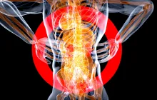
The hallmark feature of many kidney stones is colicky pain that begins in the flank or lower back and then radiates toward the groin, testicular region in men, or labial area in women. The pain might start with a dull ache and evolve into a sharp, stabbing sensation that comes in waves as the stone moves, a pattern often described as cramping. The timing of these waves can vary; some people experience brief bouts that last a few minutes, while others endure longer episodes that persist for hours. The precise location of repatterned pain often shifts as the stone advances through the ureter, sometimes prompting patients to change positions or pace the room in search of relief. In addition to back and groin pain, some individuals feel pain in the abdomen or experience a sensation of fullness that seems out of proportion to what is seen on exams. The intensity of pain does not always correlate with stone size—a small stone in a sensitive segment of the ureter can cause intense pain, while a larger stone situated in a less reactive segment might cause less dramatic symptoms. Understanding this variability helps patients and clinicians approach evaluation with an open mind and avoid premature conclusions about severity based solely on how strong the pain feels.
Urinary changes and signs to monitor
Urinary changes are common and informative when assessing possible kidney stones. Blood in the urine, described as hematuria, may be visible to the naked eye or detectable only under a microscope, indicating that stone movement is causing microtrauma to the urinary tract lining. Urine color may appear pink, red, or brown, and some individuals notice cloudiness or a foul odor as infection or mineral content shifts accompany stone passage. A frequent urge to urinate, a burning sensation during urination, or the sensation of not fully emptying the bladder can also accompany stone movement, especially as the stone approaches the bladder. In some cases, people experience reduced urine output or the sudden onset of difficulty passing urine, which signals a potential obstruction in the urinary tract that requires prompt medical attention. Keeping track of whether there is steady blood in the urine, persistent urgency, or unusual discharge can help differentiate stone-related symptoms from routine urinary infections or other conditions that mimic stones. Hydration status and the clarity of one’s urinary stream also provide valuable clues to clinicians who are evaluating symptoms in the context of kidney stone risk.
Systemic signs and warning indicators
Beyond localized pain and urinary changes, some individuals develop systemic signs that reflect the body’s response to irritation, dehydration, or infection. Nausea and vomiting can accompany intense renal colic and may persist for hours or days, sometimes complicating attempts to drink fluids. Sweating, paleness, and a sense of lightheadedness are other systemic responses that can accompany severe pain, particularly when the pain is persistent or unrelieved. Infections associated with kidney stones, such as pyelonephritis or lower tract infections, may present with fever and chills in addition to the urinary and pain symptoms. These systemic features are especially important to recognize because they can indicate a more serious problem that requires urgent evaluation and management. Individuals who notice fever with flank pain or fever with bloody urine should seek medical care promptly, as infection can rapidly worsen and lead to complications if not treated in a timely manner.
Distinguishing kidney stones from similar conditions
Several conditions mimic the pain and urinary symptoms of kidney stones, including urinary tract infections, appendicitis, gallbladder disease, diverticulitis, pancreatitis, and problems related to the reproductive organs. The pain location, character, and associated symptoms often help differentiate these conditions, but overlapping features can occur. For example, a urinary tract infection might cause burning on urination and fever with mild flank discomfort, while appendicitis may present with less predictable pain progression and abdominal tenderness rather than classic colicky renal pain. Medical professionals use a combination of history, physical examination, laboratory testing, and imaging to distinguish kidney stones from other disorders. Persistently severe pain, the presence of fever with flank symptoms, or signs of dehydration that do not improve with fluids should prompt urgent assessment to avoid missing a potentially serious alternative diagnosis. Patients should communicate all symptoms clearly, including their onset, progression, and any prior stone history, to support accurate evaluation and timely treatment decisions.
Risk groups and variability in symptoms across populations
While kidney stones can affect adults of any age, certain groups demonstrate higher incidence and sometimes different symptom patterns. Men are more frequently affected than women, and adults between the ages of roughly twenty and sixty-five show higher risk compared with other age groups. People living in hot climates or those who engage in strenuous physical activity may be more prone to dehydration, which concentrates minerals in the urine and raises stone risk. Individuals with a family history of stones, certain metabolic disorders such as hypercalciuria, gout, or cystinuria, and those with radiotherapy or some medications that influence mineral balance can have a higher likelihood of stone formation. Some populations may experience stones at younger ages due to genetic predispositions, while others present later due to accumulated risk factors over time. The symptom profile may vary with stone size and surface characteristics; a very small stone may pass with minimal or intermittent pain in some people, whereas a larger stone can produce abrupt, severe colic. Recognizing this variability helps clinicians tailor diagnostic and therapeutic approaches to each patient’s unique situation.
Urgent care red flags and when to seek immediate help
There are clear warning signs that should prompt immediate medical attention, even if pain seems to improve temporarily. Severe, unrelenting pain that prevents normal function, accompanied by vomiting and inability to keep fluids down, can lead to dehydration and may require intravenous fluids and pain relief in a clinical setting. A fever above 38.0 C (100.4 F) with flank pain or urinary symptoms raises concern for infection, which can progress rapidly if not treated. The appearance of shaking chills, fainting, confusion, or a sudden drop in blood pressure is a medical emergency that warrants urgent care. In pregnant individuals, any intense flank or abdominal pain should be assessed promptly because pregnancy introduces unique considerations for both the mother and the fetus. Additionally, signs of urinary obstruction with anuria or severely reduced output can signal that urine is not passing properly and requires urgent evaluation to prevent kidney damage. In all these scenarios, seeking prompt evaluation minimizes risk and often leads to faster relief and safer management.
Diagnostic approaches doctors use to evaluate kidney stone symptoms
When kidney stones are suspected, clinicians combine clinical information with targeted tests to confirm the diagnosis and guide treatment. A noncontrast helical CT scan of the abdomen and pelvis is a highly sensitive and specific test for detecting most kidney stones and can reveal their size, location, and potential complications. Ultrasound is a radiation-free option that is particularly useful in certain populations, such as pregnant individuals and children, or when radiation exposure is a concern. A plain X-ray of the kidneys, ureters, and bladder (KUB) may identify radiopaque stones but is less sensitive than CT for many stone types. Urinalysis helps detect microscopic or visible blood in the urine, signs of infection, and crystals that can inform stone type and the need for specific treatment. Blood tests can assess kidney function and metabolic contributors, including electrolyte balance and calcium or uric acid levels, which influence both diagnosis and prevention strategies. The combination of imaging and laboratory tests provides a comprehensive picture that supports accurate treatment decisions and helps predict the likelihood of spontaneous stone passage versus the need for intervention.
Managing symptoms while awaiting diagnosis or treatment
While awaiting a definitive diagnosis or in the early stages of stone evaluation, several measures can help reduce discomfort and protect kidney function. Adequate hydration is essential, but it should be approached gradually and under medical guidance if there is significant pain or vomiting. Clear fluids such as water or oral rehydration solutions can help maintain urine production and dilute minerals that contribute to stone formation, though excessive intake in the presence of a severe obstruction or infection may be inappropriate; clinicians tailor fluid recommendations to each patient’s situation. Analgesia is often necessary, with nonsteroidal anti-inflammatory drugs like ibuprofen or acetaminophen commonly used, depending on kidney function and other health considerations. In some cases, doctors may prescribe medical expulsive therapy to facilitate stone passage, particularly for stones in specific locations and of a size that is likely to pass spontaneously. This therapy, when appropriate, can shorten the time to passage and reduce the need for invasive intervention. It is crucial to follow medical advice and avoid self-prescribing medications that could interact with existing health conditions, and to seek urgent care if pain becomes uncontrolled or signs of infection emerge during home management.
Treatment pathways after evaluation: what to expect
Treatment decisions depend on several factors, including stone size, shape, composition, location within the urinary tract, the patient’s symptoms, and overall kidney function. Smaller stones that are likely to pass on their own may be managed with analgesia, hydration, and timed observation, along with guidance on when to return for reassessment. For stones that are less likely to pass naturally, or for those associated with persistent obstruction or infection, doctors may pursue active interventions. Medical expulsive therapy can be effective for certain distal ureteral stones by relaxing the smooth muscle in the ureter and easing stone passage. If stones are larger, located unfavorably, or associated with obstruction, procedures such as extracorporeal shock wave lithotripsy to break the stone into fragments, ureteroscopy to retrieve or break the stone via a scope, or percutaneous nephrolithotomy for larger stones within the kidney may be recommended. Each approach carries its own risks, recovery times, and success rates, and the choice is guided by the stone’s characteristics and the patient’s health status. Ongoing follow-up is important to confirm resolution, monitor for recurrence, and manage any residual fragments that could cause future trouble.
Prevention and long-term management to reduce recurrence
Preventing kidney stone recurrence involves a combination of lifestyle adjustments, dietary modifications, and, in some cases, medication tailored to the stone type. Increasing daily fluid intake to maintain a high urine volume is one of the most effective strategies and should be sustained as a long-term habit; consistency matters as spacing out intake can allow minerals to re-concentrate at times of lower hydration. Dietary changes may include balancing salt and animal protein intake, moderating foods rich in oxalates if a person forms calcium oxalate stones, and ensuring adequate calcium intake through foods rather than supplements unless advised by a clinician. For certain metabolic conditions, doctors may prescribe medications that alter urine chemistry—such as thiazide diuretics for hypercalciuria or allopurinol for uric acid stones—to reduce the likelihood of new stones forming. Regular follow-up with a healthcare provider is essential to monitor urinary tract health, adjust prevention strategies, and identify any metabolic risks that require intervention. Advocating for healthy hydration, a balanced diet, and timely medical evaluation creates a proactive framework for minimizing future stone formation and supporting overall kidney health.